
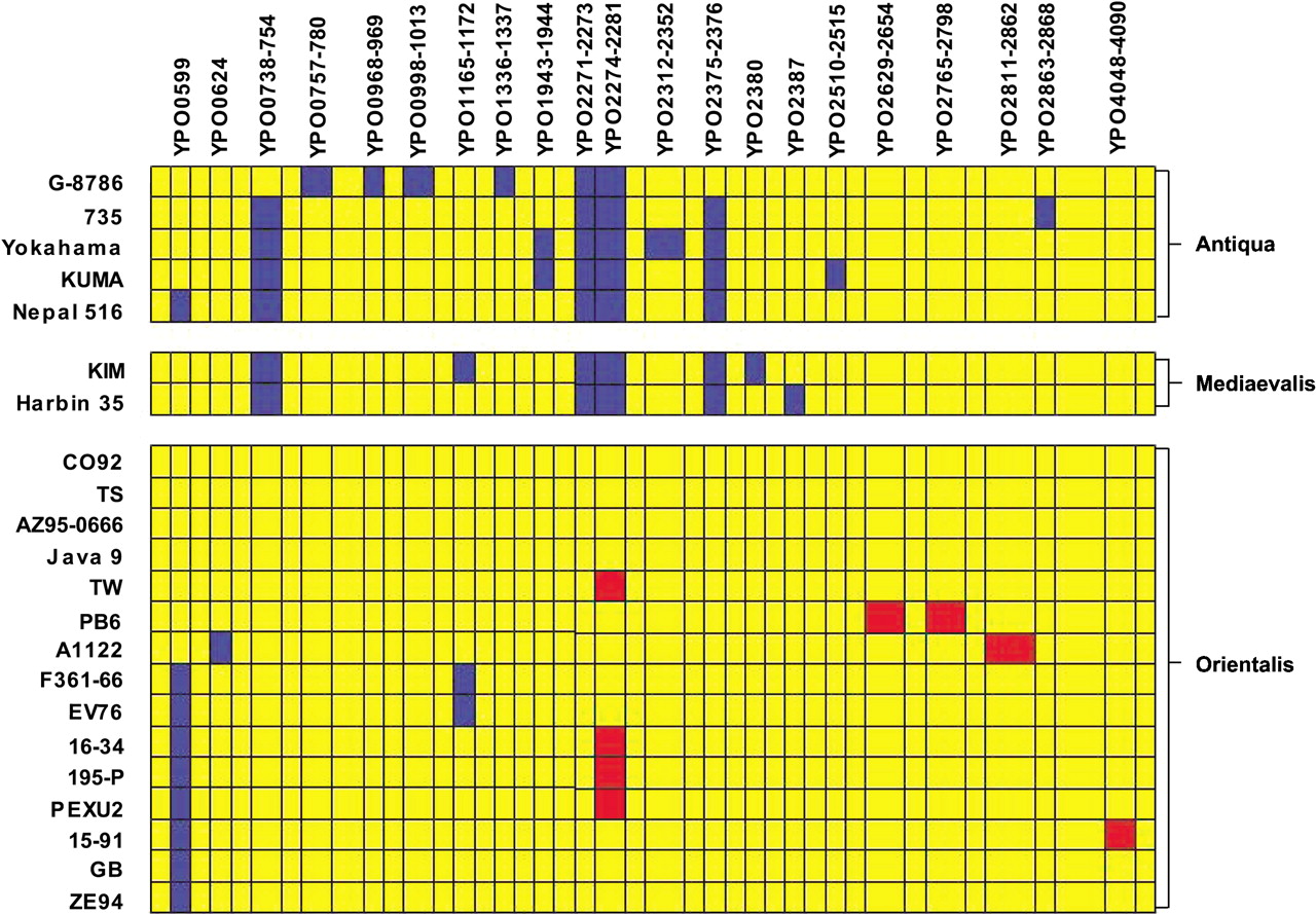
Figure 2
Schematic of chromosomal comparison of 22 strains of Y. pestis detailing all of the regions of divergence from CO-92. Strains are grouped by biovar. The 102-kb unstable region (YPO1902–YPO1967) has not been included in this comparison. Gene status is color coded as in Figure 1 for ease of comparison, with yellow indicating presence, blue indicating absence, and red indicating a duplication.


