
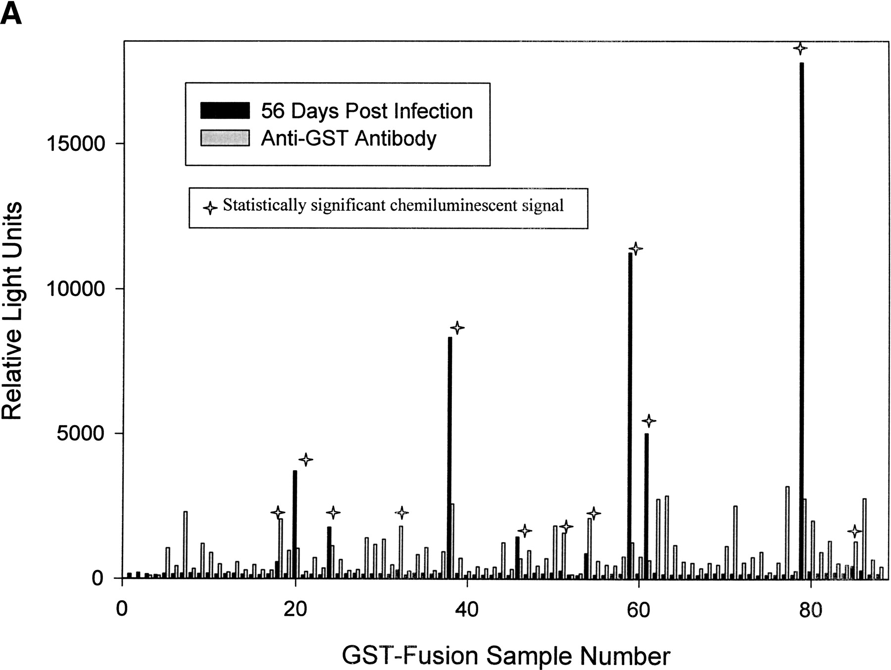

(A) Comparison of the chemiluminescent signal generated from exposing immobilized GST-fusion proteins to either anti-GST-HRP or serum from rabbits 56 d after Treponema pallidum infection. As described in Fig. 2A, GST-fusion proteins from crude Escherichia coli extracts were selectively immobilized on glutathione-coated 96-well plates. To compare relative amounts of immobilized GST-fusion protein, anti-GST-HRP antibody was incubated in the wells instead of rabbit serum. A plate reader monitored chemiluminescence and the mean relative signal is presented next to the chemiluminescent signal measured in the immunoassay technique (Fig. 2B). A star has been placed above each of the 12 samples with a statistically significant signal (Table 2). (B) A scatter plot presents the absence of a correlation between GST-fusion protein amount and antigenic properties. The mean relative chemiluminescent signal detected from captured anti-GST-HRP antibody is plotted along the X-axis. The mean relative chemiluminescent signal detected from captured IgG antibodies present in rabbit serum 56 d after T. pallidum infection is plotted along the Y-axis in log scale. Samples with a statistically significant signal have been labeled (Table 2).


