
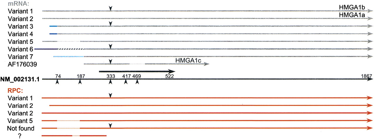
Figure 6.
HMGA1 RPCs aligned with mRNA splice isoforms. (Black arrow in the middle) The NM_002131.1 RefSeq mRNA; (thick black arrow) its CDS position. Above are transcript variants in gray, below are RPCs in red; different shades of blue lines represent different alternative exons, and the hatched line in variant 6 represents an Alu element. (Downward-pointing arrowheads) The position of an additional 33 bp between exons 3 and 4 in some of the transcripts and RPCs. (Upward-pointing arrowheads) Splice positions; (dotted lines) deletions or exon skipping.


