
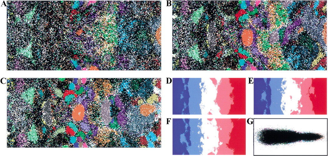
Figure 2.
SOMs for 1-kb sequences of 65 bacterial genomes. (A,B,C) Di-, tri-, and tetra-SOMs, respectively. Lattices are colored as described in Fig. 1,A–C. (D,E,F) G+C% for each weight vector is shown as described in Fig. 1, D–F. (G) Classification by the initial weight vectors for the di-SOM.


