
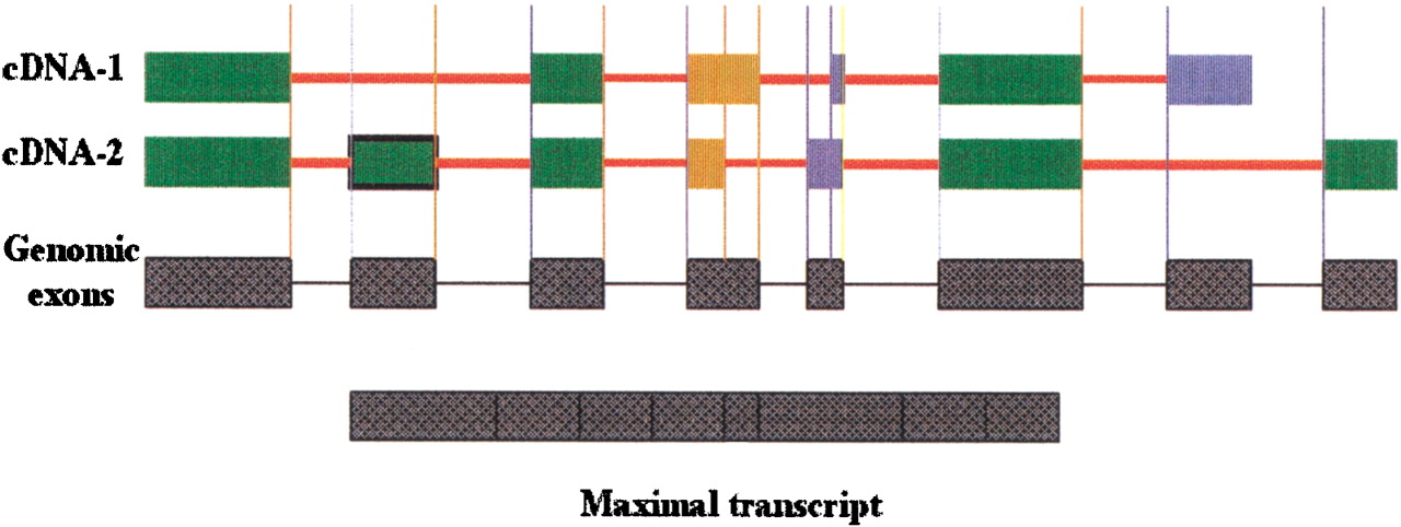
Figure 7.
Types of splice variants. Vertical lines indicate splice sites, blue for 3′ splice sites and gold for 5′ splice sites. Genomic exons are shown at bottom. Exons are colored to indicate type of variation as follows: green exons have conserved splice sites; blue exons have multiple splice sites at the 5′ end; gold exons have multiple splice sites at the 3′ end. Cryptic exons are shown with a black frame. Terminal exons occurring in the intron of another clone are shown in blue.


