
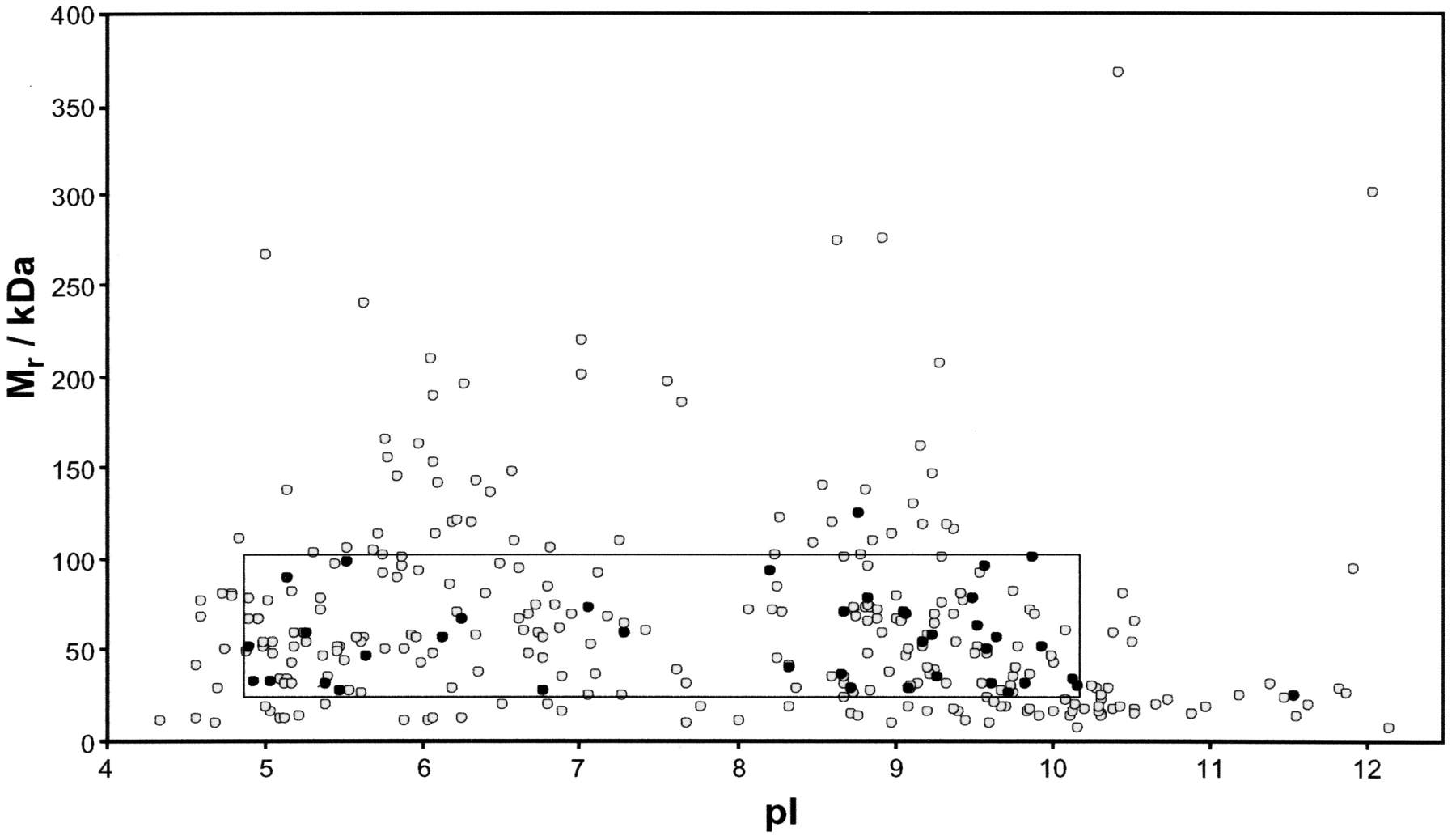
Figure 4.
Virtual 2D gel of proteins identified in the spliceosome preparation. The coordinates are the theoretical molecular mass and isoelectric point for each protein. The gray circles represent factors identified in this study, and the black circles represent proteins identified in this and our previous study (Neubauer et al. 1998). The box indicates the coordinate space spanned by our previous investigation using 2D gel electrophoresis.


