
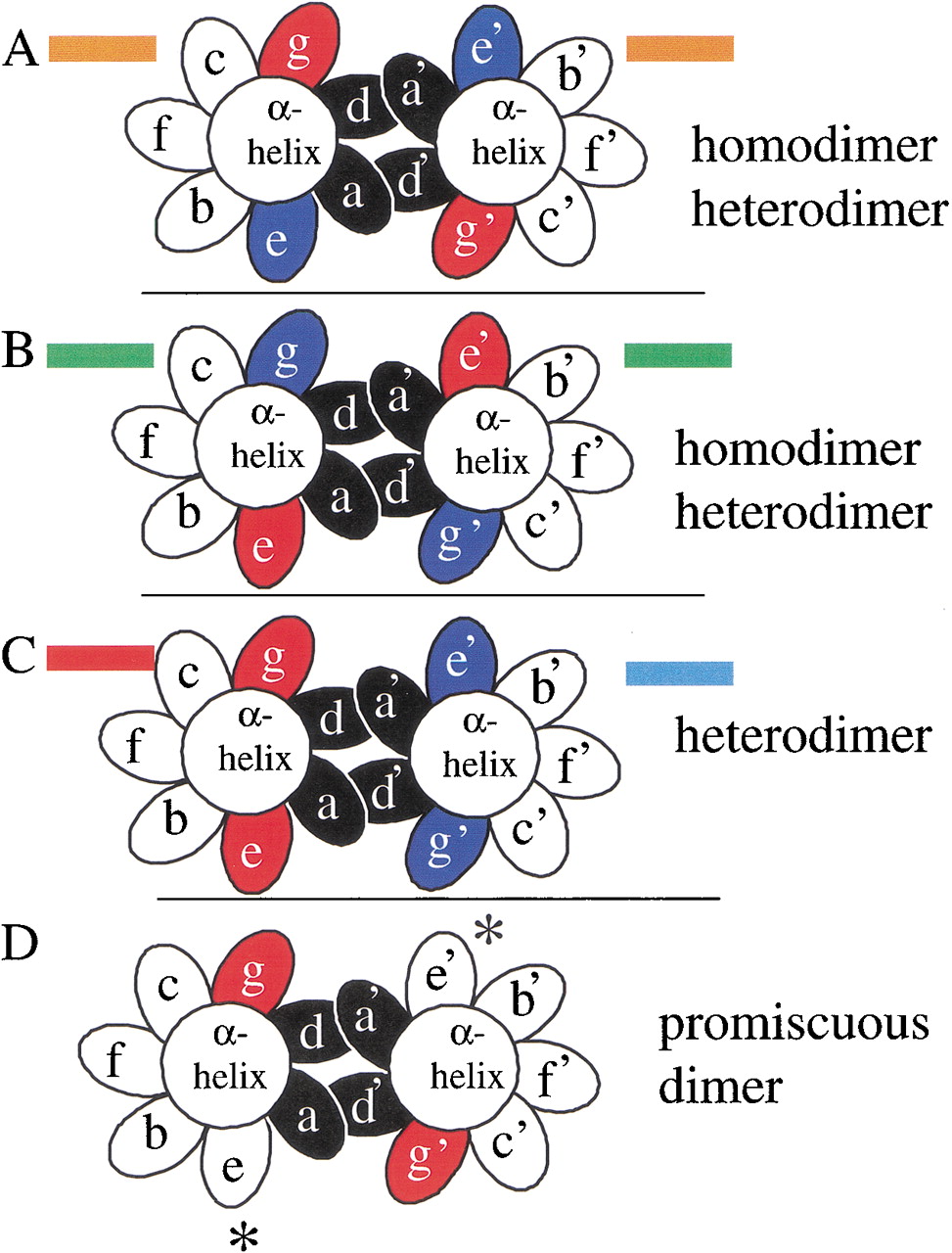
End view, looking from N terminus to C terminus, of a coiled coil with the seven unique positions of the heptad presented as ellipses. The ‘a’ and ‘d’ positions are colored black. The four possible combinations of acidic and basic amino acids in the ‘g’ and ‘e’ positions are presented and color coded as used in Figure 2. (A) An α-helix with a g↔e‘ pair containing an acidic amino acid in the ‘g’ position and a basic amino acid in the following ‘e’ position (orange in Fig. 2) can form a homodimer or heterodimer with a similarly charged α-helix. (B) An α-helix with a g↔e‘ pair containing a basic amino acid in the ‘g’ position and an acidic amino acid in the following ‘e’ position (green in Fig. 2) can form a homodimer or heterodimer with a similarly charged α-helix. (C) A heterodimer between an acidic g↔e‘ pair (red in Fig. 2) and a basic g↔e‘pair (blue in Fig. 2). (D) A dimer with an “incomplete”g↔e‘ pair resulting in promiscuous dimerization.


