
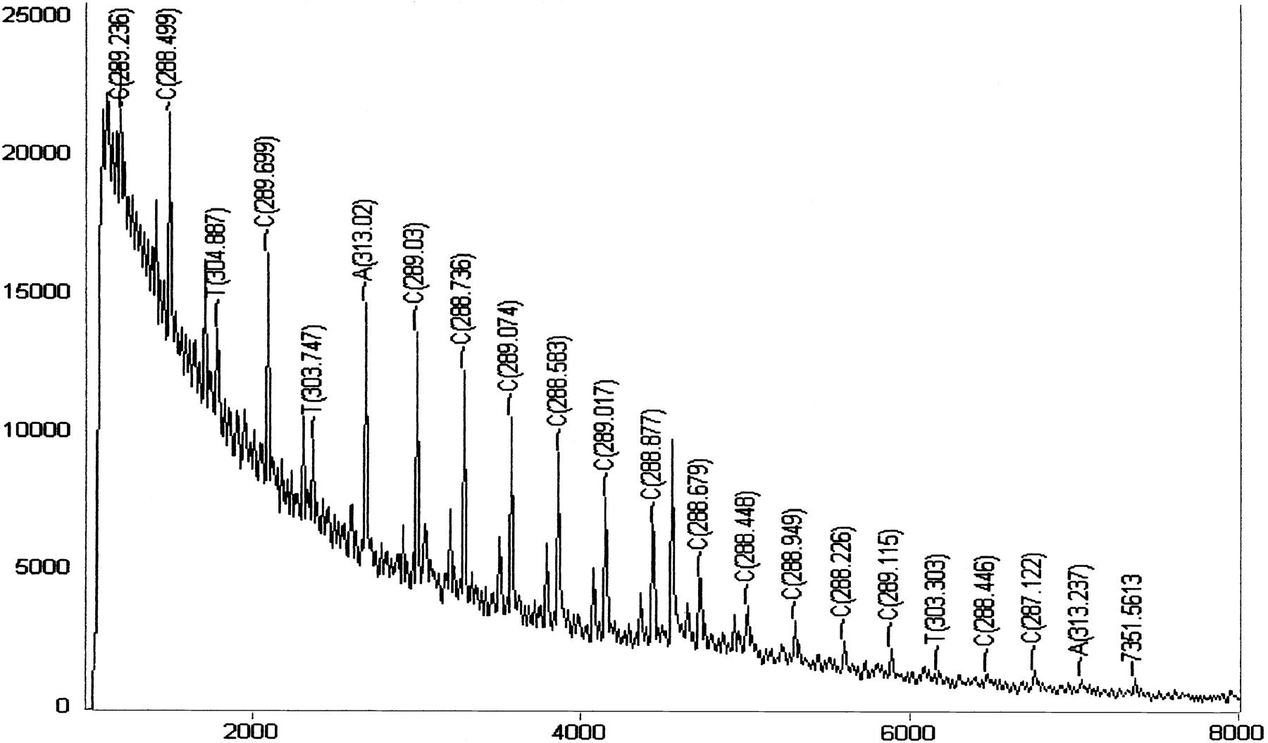
Figure 3.
Negative-ion MALDI-TOF-MS spectrum of a PCR product after exonuclease digestion. This spectrum clearly shows the presence of 11 consecutive cytidines flanked by the known sequence at the 3′ and 5′ ends. The doubly charged ion of the remaining undigested PCR product gave an approximate mass-to-charge (m/z) value of 4581 between the sixth and the seventh sequential cytidines. The secondary series of peaks visible between 2000 and 4500 amu are mainly caused by partial digestion of the complementary strand of the PCR product from the 3′ end.


