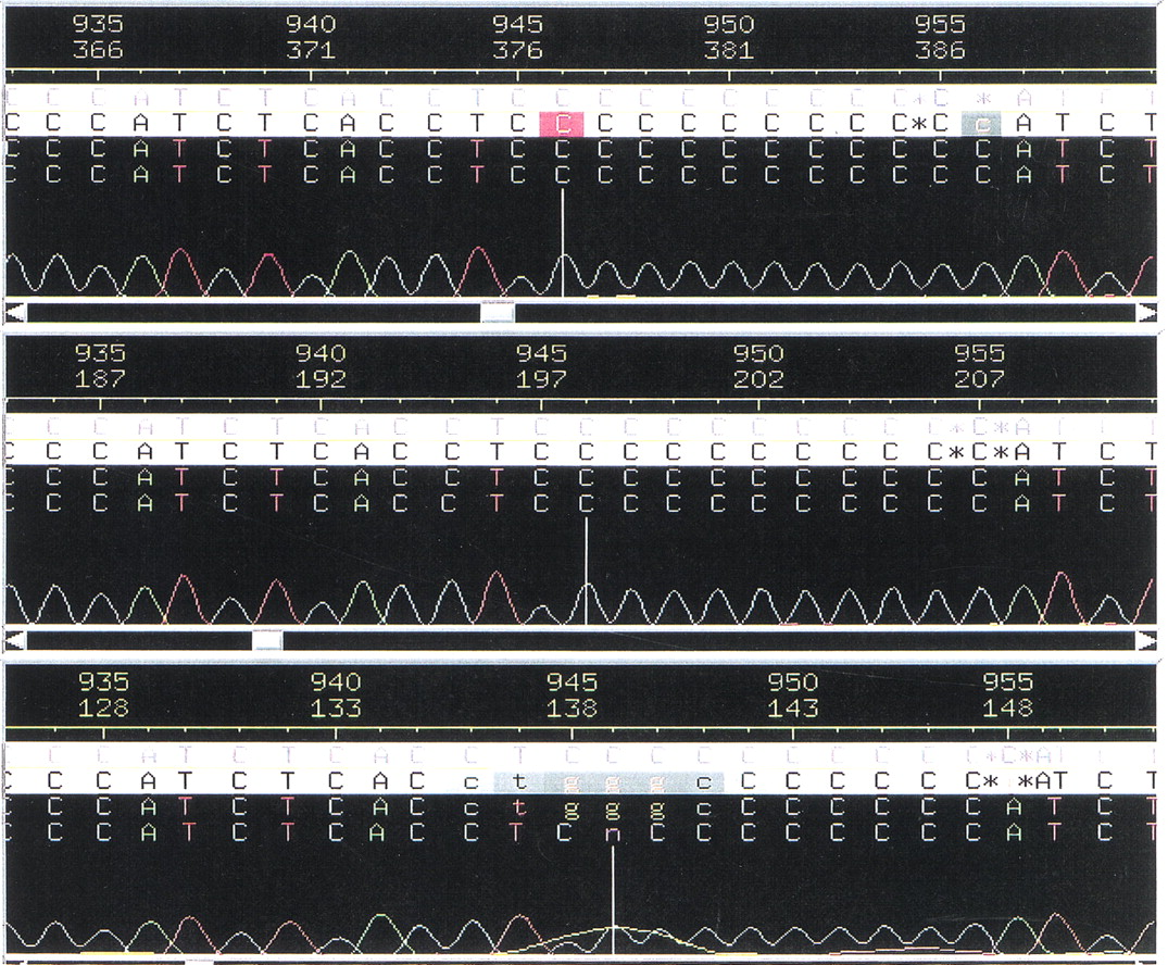
Validation of DNA Sequences Using Mass Spectrometry Coupled with Nucleoside Mass Tagging

(Downloading may take up to 30 seconds. If the slide opens in your browser, select File -> Save As to save it.)

Click on image to view larger version.

Figure 1.
Figure 1.

Three fluorescent gel electrophoresis sequencing chromatograms of the same genomic DNA region from chromosome 19 showing the unresolved region of the polycytosine string. Each of these three chromatograms indicated a different number of cytidines.

This Article

  1. Genome Res. 12: 1135-1141

Preprint Server