
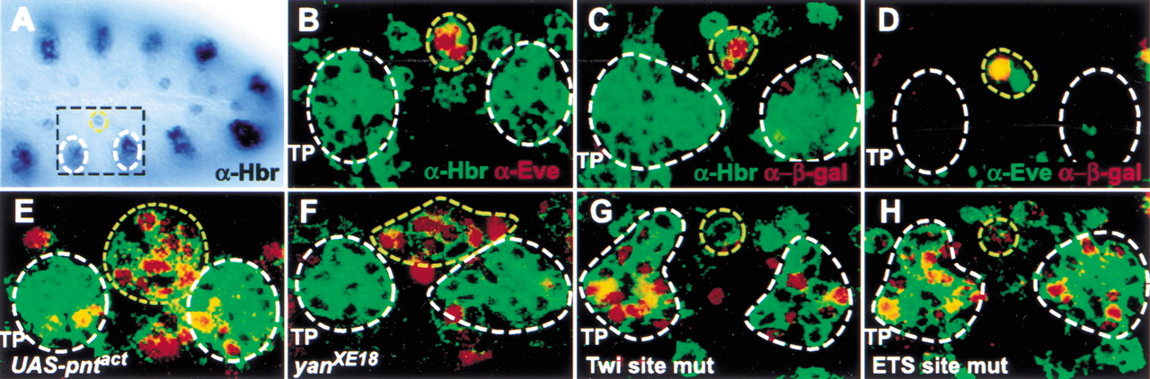
The Hbr DME. In all panels, white circles mark the developing tracheal cells, and a yellow circle denotes the mesodermal progenitors. (A) Stage 11 embryo stained with antibodies against Hbr. Expression can be seen in the ectodermally derived tracheal pits and the mesodermal progenitors. The portion of each hemisegment shown in the remaining panels is indicated by a black box. (B) Double-labeling for Hbr (green) and Eve (red) shows that the two proteins are found in the same cells in the dorsal mesoderm. Note that Hbr is membrane-associated, whereas Eve is nuclear. Additional mesodermal cells expressing Hbr but not Eve can be seen on both sides of the Eve progenitors. (C) The Hbr DME reporter (nuclear β-galactosidase, red) is expressed in a subset of the Hbr-positive mesodermal progenitors (green). The cells of the developing trachea do not express the reporter. (D) Double-labeling for Eve (green) and the DME reporter (red) show that the DME is expressed in at least one of the Eve progenitors. (E,F) Ectopic activation of the Ras/MAPK pathway through either mesodermal expression of activated Pnt (E) or loss-of-function of the repressor Yan (F) is accompanied by an expanded number of both Hbr- (green) and DME- (red) expressing cells. (G) Mutation of the Twi binding sites in the DME results in a loss of mesodermal reporter gene activity and the acquisition of expression in the developing trachea. (H) Mutation of the Ets binding sites in the DME causes the acquisition of expression in the developing trachea but has only a minimal effect on mesodermal expression. TP, tracheal pits. All panels show anterior to the left and dorsal up.


