
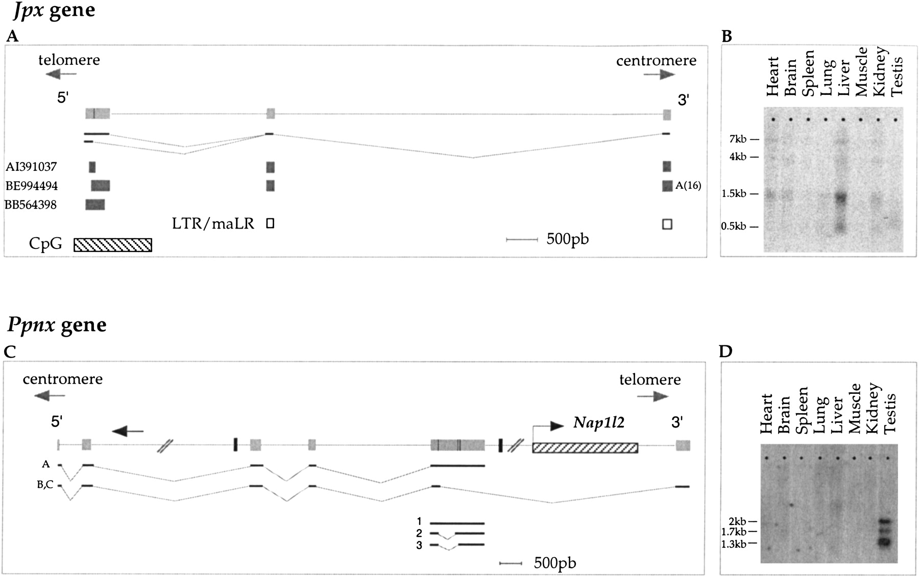
Cloning and characterization of Ppnx and Jpx genes. (A) Jpx gene. Schematic diagram showing the three exons (light grey boxes) of the murine Jpx gene, at positions 343745–343379 (exon 1), 340888–340796 (exon 2), 334465–334364 (exon 3), as defined by the three ESTs represented as dark grey boxes above the map. ESTs are identified by their GenBank accession number. The presence of an alternative splice at position 343626 nt in exon 1 generates either a 315- or 562-nt transcript. The presence of a poly(A) tail in EST BE994494 identifies the 3′ end of the gene. The first exon may correspond to the 5′ end of the gene, as a CpG island (diagonally hatched box) lies in the vicinity. The two last exons are LTR/MaLR-repeat elements (white boxes). (B) Northern analysis using a PCR probe corresponding to exon 1 of the Jpx gene after 5 d exposure. (C) Ppnx gene. Schematic diagram illustrating the six exons of the murine Ppnx gene. All exons and introns are represented to scale with the exception of introns 2 and 5 that are very large (21707 bp and >45389 bp, respectively). Each exon (grey) is identified by its coordinates: exon 1, 591535–591619; exon 2, 592146–592314; exon 3, 614021–614268; exon 4, 615349–615488; exon 5, 618116–>619619 (with three alternative splice donor sites at position 618262, 618719, and 618758), and exon 6, 665008–665306. The black boxes represent two pseudogenes predicted byGENSCAN; the small black arrow shows an antisense transcription detected in intron 2. The single-exon NapIl2gene is shown diagonally hatched. Three independent clones A, B, and C obtained after screening a testis cDNA library are shown below the genomic structure. Clone A does not contain either a poly(A) consensus signal or a poly(A) track and has an in-frame stop codon present in its most 3′ part. The end of the 3′ UTR track is likely 300 bp downstream of the stop codon (unpubl.). Clones B and C possess a stop codon in exon 6 followed by a 3′ UTR of 177 bp, containing both a poly(A) signal and a poly(A) tail. Three of the RT-PCR products suggesting the existence of alternative splice donor sites in exon 5 are also represented (1, 2, and 3). (D) Northern analysis ofPpnx expression. A mouse multiple tissue Northern blot was hybridized with a probe corresponding to the third exon ofPpnx. The Ppnx hybridization signals were obtained after 8 d exposure.


