
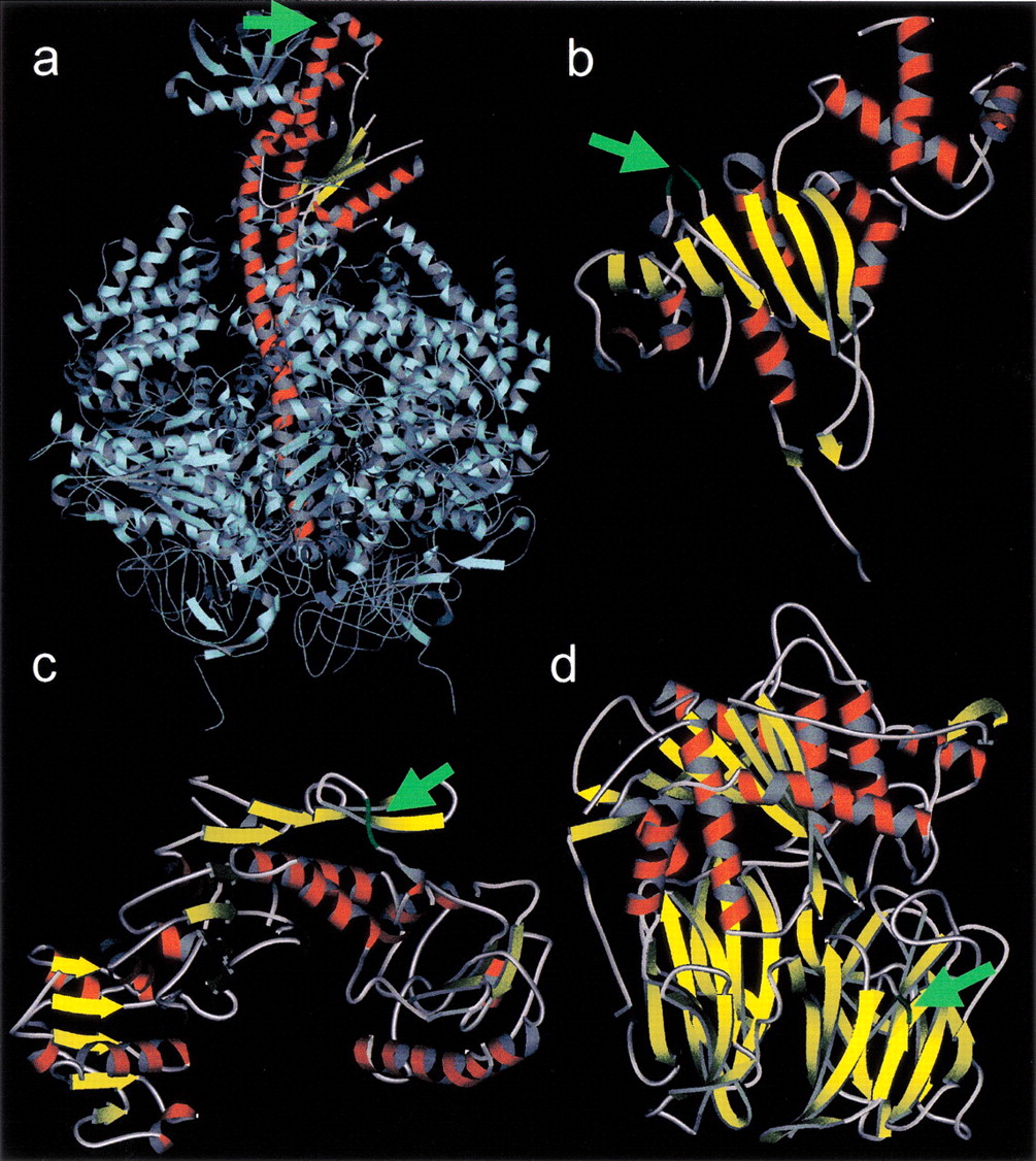
Figure 3.
Predicted insertion sites of the RPE-2 (a, b) and RPE-3 (c, d). Green segments labeled by the green arrows in the reference structures indicate the insertion sites. (a) Bovine mitochondrial ATP synthase. γ chain is shown in red. (b) Streptococcus pneumoniae rRNA methyltransferase used as a reference for the R. conoriidimethyladonosine transferase. (c) E. coliUDP-N-acetylmuramoylalanine–D-glutamate ligase. (d) Pig prolyl oligopeptidase used as a reference for the R. conoriiprotease II. Seven cases corresponding to the predicted insertions sites for the RPE-1 have been previously reported (Ogata et al. 2000). The images are generated with MolScript(http://www.avatar.se/molscript/).


