
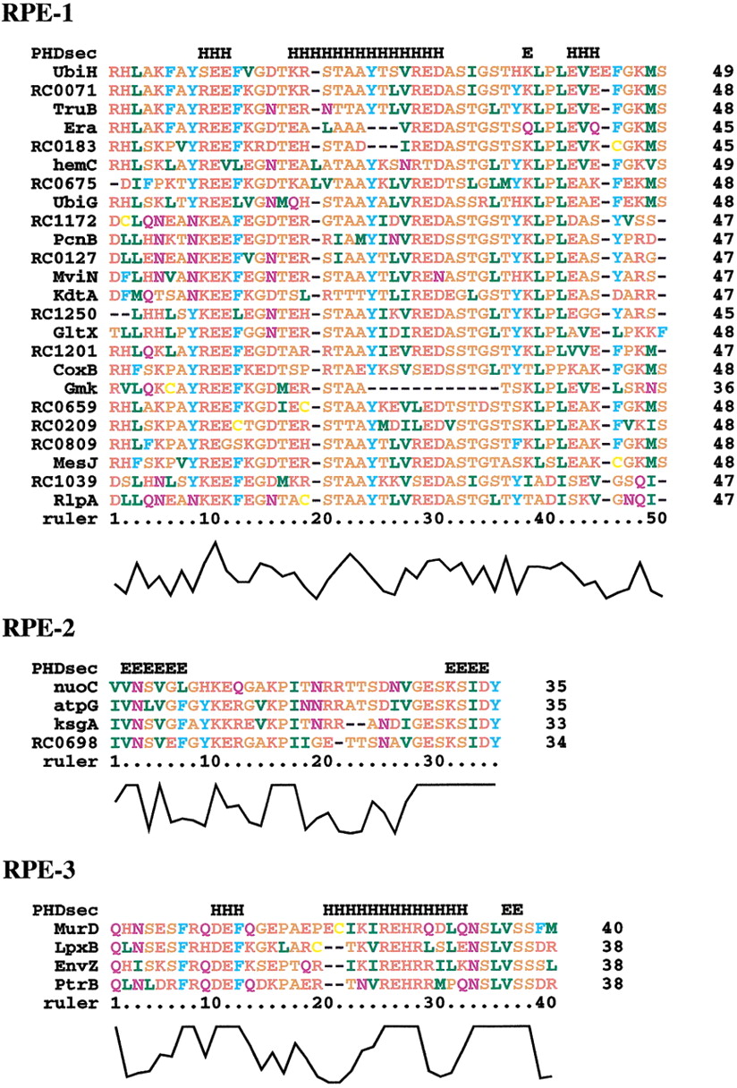
Figure 2.
Multiple sequence alignments for the amino acid sequences translated from the palindromic sequences (RPE-1, RPE-2, and RPE-3). Amino acid residues are colored as follows: blue for F, W, and Y; yellow for C; orange for A, G, P, S, and T; green for I, L, M, and V; red for D, E, H, K, and R; purple for N and Q. The letters ‘H’ and ‘E’ in the first line of each alignment represent the predicted α-helices and β-strands, respectively. ClustalX alignment quality scores are shown at the bottom of the alignment.


