
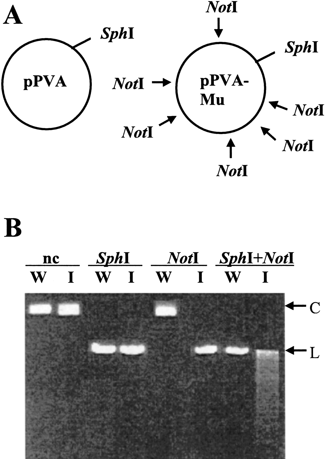
Figure 3.
Restriction analysis of pPVA and pPVA-Mu plasmid pools. (A) Plasmid map of pPVA and predicted map of pPVA-Mu are shown. Arrows indicate inserted NotI sites in different molecules. (B) Plasmids were digested with enzymes and analyzed by electrophoresis on a 1.8% TBE gel parallel to nondigested (nc) plasmids. pPVA and pPVA-Mu are denoted as ‘W’ and ‘I,’ respectively. Migration of circular (C) and linear (L) plasmid forms are indicated.


