
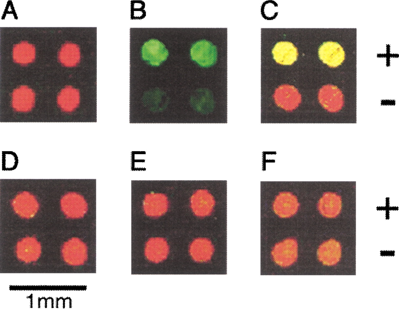
Detecting protein–DNA interactions. (A) Two types of DNA labeled with Cy5 (650/670 nm) were spotted on a glass slide (represented as false-colored red), one containing a Fos–Jun bound sequence (+) and the other not (−). (B) The sequence-specific binding of the complex of the Jun labeled with TAMRA (542/568 nm) and unlabeled Fos to the DNA (+) was detected as false-colored green spots. (C) The same result as in B is represented as a superposition of red and green color. (D–F) As negative controls, the solution of TAMRA-labeled Jun (D) without Fos, (E) with unlabeled Fos, and an excess of competitive DNA, and (F) with unlabeled Jun were probed, and no binding was observed (shown by the superposition of red and green color).


