
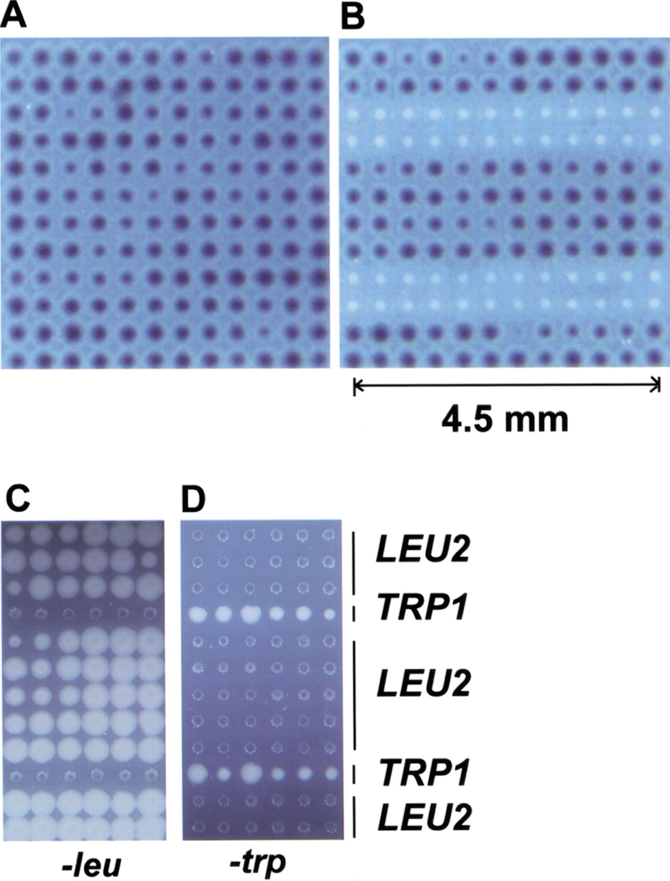
Cell microarrays of E. coli and S. cerevisiae. (A) E. coli cell microarrays (144 colonies) expressing β-galactosidase in the presence of S-gal (3,4-cyclohexenoesculectin-β-D-galactopyranoside), a chromogenic substrate for β-galactosidase. (B) E. coli cell microarrays with 48 colonies that did not express β-galactosidase. (C) S. cerevisiae microarrays grown on synthetic medium without leucine. (D) S. cerevisiae microarrays grown on synthetic medium without tryptophan. Bacterial cell microarrays were placed on the surface of LB in the presence of 50 μg/mL S-gal at 37°C overnight. Yeast cell microarrays were placed on the surface of dropout media indicated in the figure at 30°C overnight.


