
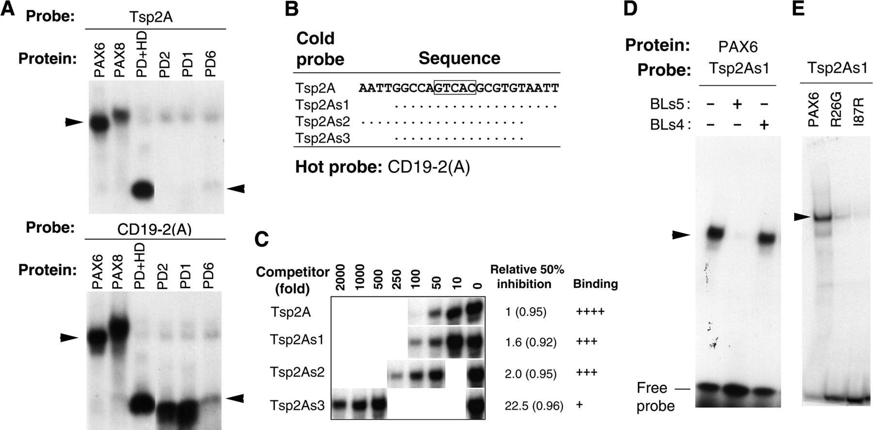
(A)EMSA of PAX6-specific binding site Tsp2A with whole or partial Pax proteins (the abbreviation for proteins are defined in the legend for Fig. 1). CD19-2(A) was used as a positive control. (B) Probe sequences. Additional random sequences (GAAGAGCTC at the 5′ ends and GAGCTCGAC at the 3′ ends) were added to ends of the probe sequences shown here as possible docking sites. The relative binding affinities were deduced based on the amount of competitor inhibiting protein binding by 50% from EMSA in panelC. The core sequence GTCAC for binding by the N-terminal subdomain of the PD (Jun and Desplan 1996) is boxed. Dots represent nucleotides identical to those in Tsp2A. The numbers in parentheses are the correlation coefficients (R) between the amount of cold probe and the amount of the complex formed. (D) Sequence-specific binding of PAX6 to Tsp2As1. BLs5 is a PAX6-specific binding probe and BLs4 is a non-PAX6-specific binding probe (Zhou et al. 2000). (E) EMSA of PAX6, and mutant PAX6 R26G and I87R to Tsp2As1.


