
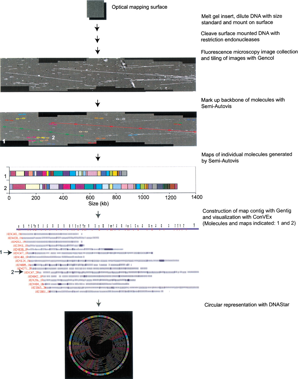
Scheme for shotgun optical mapping. High-molecular weight DNA is simply extracted from cells and deposited onto an optical mapping surface. After restriction endonuclease digestion and staining with a fluorescent dye, individual molecules are imaged by fluorescence microscopy. Images are collected using Gencol, which accumulates overlapping images in a semiautomated fashion and preserves registration. Semi-Autovis is then used to automatically convert image data into map files after a user selects molecules. Maps are then automatically contiged using Gentig, and the results are displayed using ConVEx. ConVExallows the user to edit contigs, view statistics, and browse molecular images. Finished maps are visualized as a circular chromosome using software from DNAStar.


