
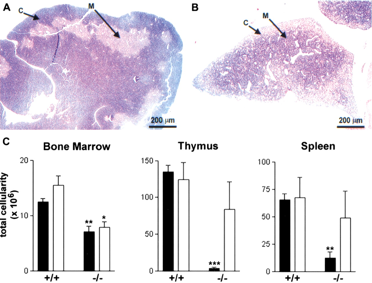
(A,B) Histopathology of 4-wk-old female (A) control and (B) Desrt −/− thymi showing the overall reduction in the size of the Desrt −/−thymus, the paler staining of the thymic cortex (C) and the darker stain of the thymic medulla (M) as compared with the control. (C) Comparison of the femoral bone marrow, thymus, and spleen cellularities of Desrt +/+ andDesrt −/−mice at 3-wk of age (solid bars) and at 6 wk (open bars). The cellularities of Desrt −/−animals were compared with the cellularity of age matchedDesrt +/+ animals and significant differences are indicated (*, p <0.05; **, p <0.01; ***,p <0.001).


