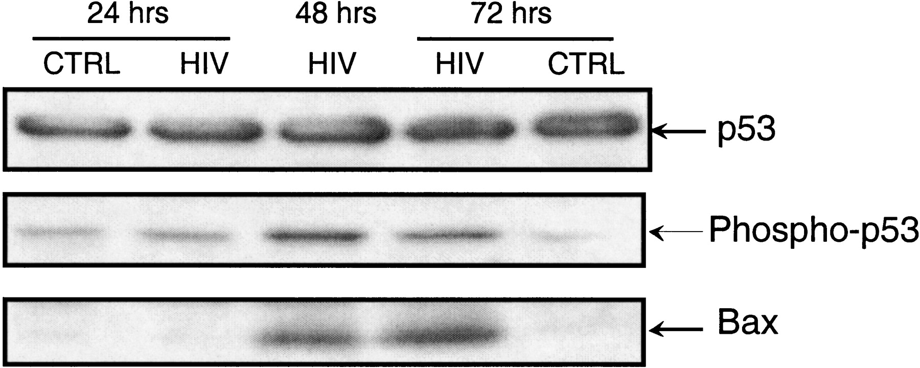
Temporal Gene Regulation During HIV-1 Infection of Human CD4+ T Cells

(Downloading may take up to 30 seconds. If the slide opens in your browser, select File -> Save As to save it.)

Click on image to view larger version.

Figure 4.
Figure 4.

Western blot for p53, phosphorylated p53, and BAX. p53 concentration remained constant but phosphorylated p53 was markedly increased in HIV 1 at 48 and 72 h, a situation mimicked for BAX.

This Article

  1. Genome Res. 11: 1198-1204

Preprint Server