
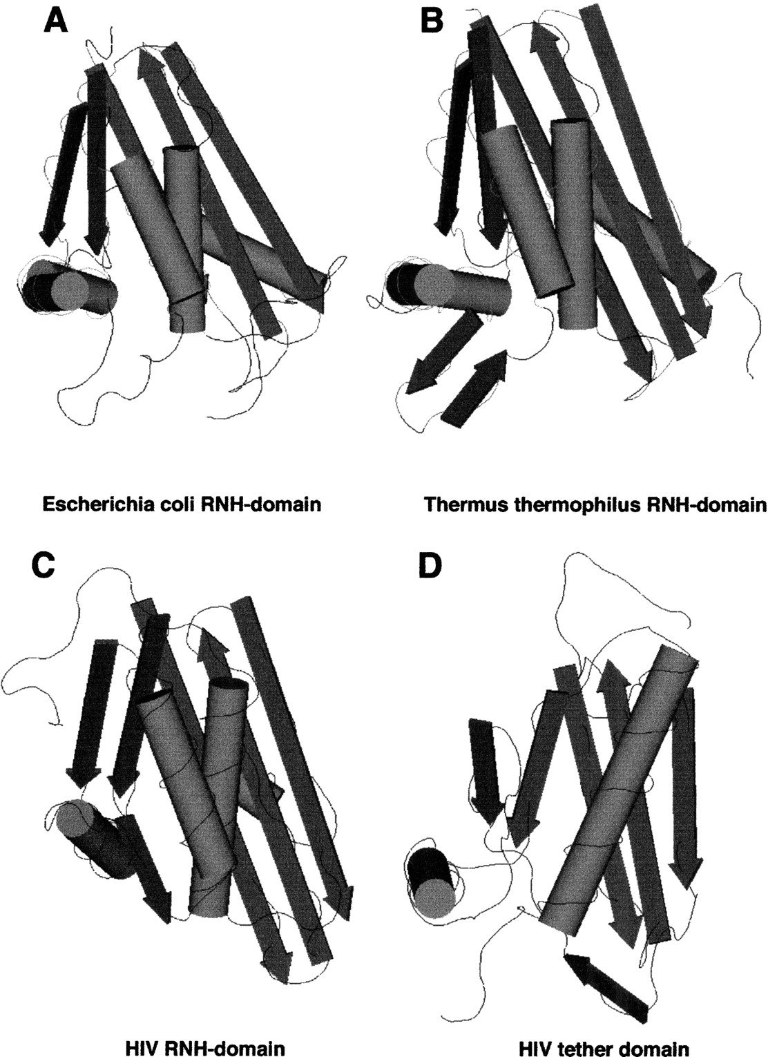
Figure 5.
Schematic three-dimensional diagrams of the RNH domains fromEscherichia coli (PDB structure 1RDD), Thermus thermophilus (1RIL) and HIV-1 (1RVT) are shown along with the tether domain of HIV-1 (1RVT). β-strands and α-helices are represented by arrows and cylinders, respectively, using theCn3D viewer software (version 3.0). Note that the tether (connection) domain has the same fold (also see Artymiuk et al. 1993) as the enzymatically active ribonuclease HI domains.


