
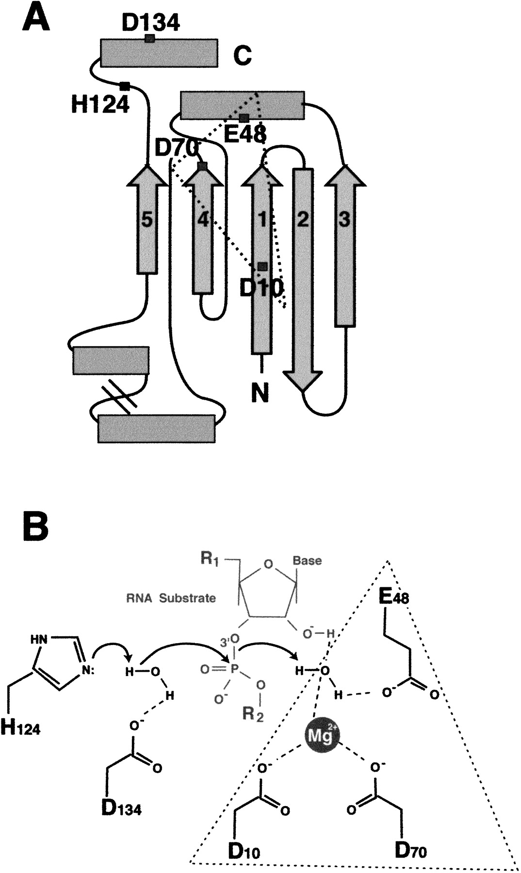
Figure 2.
(A) A simplified topological diagram of the Escherichia coli Ribonuclease HI (RNH) domain, indicating the active site residues (see Fig. 1). β-strands are indicated by arrows, and α-helices are shown by boxes. The four carboxylates and single histidine residue are shown. (B) A schematic of the proposed RNH catalytic mechanism is shown (modified with permission from Kanaya et al. 1996). The carboxylate triad typical of other endonucleases with an RNH fold (Yang and Steitz 1995; Rice et al. 1996) is indicated by the dotted triangle.


