
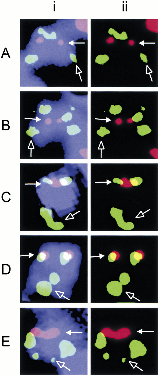
Immunofluorescence/FISH analysis of 20p12 neocentromere. Immunofluorescence was performed using CREST#6 (red) to mark the position of the neocentromere (solid arrow). FISH was performed using PACs dJ1009E24 (A), dJ811H13 (B), dJ727I10 (C), dJ742J24 (D), and dJ905G11 (E). Two sets of green FISH signals are observed on the marker chromosome invdup (20p) because of the inverted duplication of the probed segment; the signal set at the distal q′ arm is indicated by an open arrow. The combined image in (i) shows the relative positions of the signals on the marker chromosome (blue); (ii) shows only the images for the green and red signals in (i). Colocalization of the two signals appears as yellow in (ii).


