
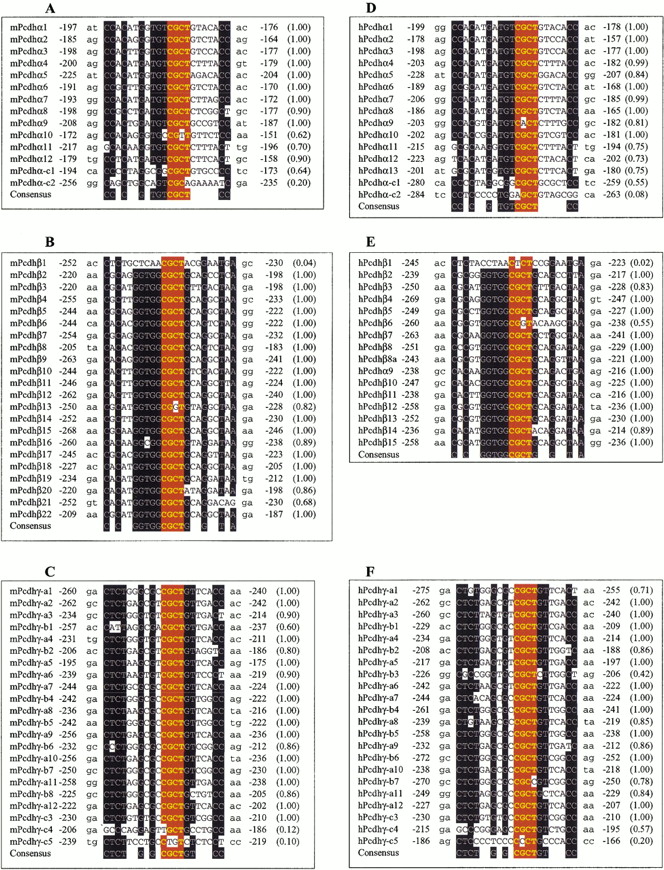
Figure 8.
Alignment of conserved sequence motif upstream of protocadherin coding region. Shown are the conserved sequences and their relative positions to the translation start codon in mouse Pcdhα (A), Pcdhβ (B), and Pcdhγ (C) and human Pcdhα (D),Pcdhβ (E), and Pcdhγ (F) gene clusters. The probability of finding the motif within -290 to -150 nucleotides upstream of the translation start codon is shown within parentheses at right. The consensus sequences are shown below each panel. The conserved nucleotides are shown with white letters on a black background. The core sequences are highlighted with yellow bold letters on a red background.


