
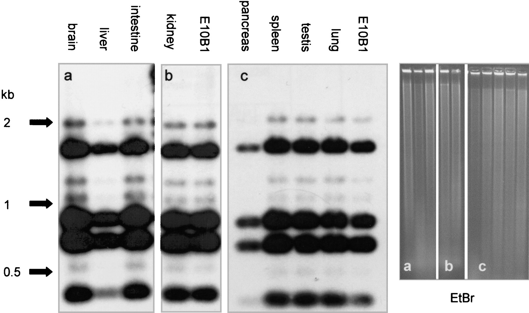
Figure 4.
Tissue distribution of the HCV. Southern blot analysis of the HCV. DNA prepared from different tissues of an HCV+ F1 mouse was digested with XbaI, size-separated, and blotted. The left panel shows hybridization with a human alphoid-2 probe. The signal obtained for the different tissues is identical to the signal obtained for the E10B1 clone. The right panel shows the ethidium bromide–stained agarose gels.


