
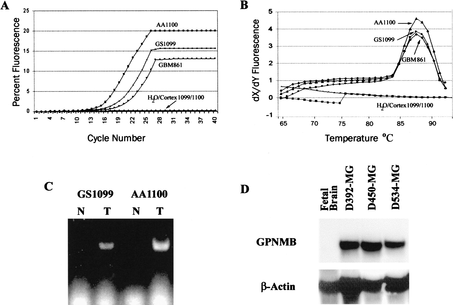
Fluorescent-PCR verification of a candidate glioblastoma marker, GPNMB. (A) Template cDNA from a bulk glioblastoma (GBM 861) and matched glioma/normal tissue pairs (GS1099/Cortex1099 and AA1100/Cortex1100) were amplified with primers specific for GPNMB. (B) Melting curve analysis is performed simultaneously to optimize detection temperature, revealing a single peak consistent with a single amplification product. (C) After fluorescent-PCR, all reaction products were visualized on an agarose gel to verify a single product of the correct size. (D) Northern blot of normal fetal brain and three established GBM cell lines also show a difference in expression forGPNMB.


