
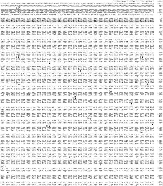
cDNA and deduced amino acid sequence of DLGR-2. This figure is compiled from the sequences of the overlapping cDNA clones 5′C-2, 5B, and 3′4-J (Fig. 1A). Nucleotides are numbered from 5′ to 3′ end, and the amino acid residues are numbered starting with the first ATG in the open reading frame. Introns are indicated by arrows and numbered 1–14. The seven membrane-spanning domains are boxed and labeled TM I—VII. The proposed signal sequence is shaded. Spades indicate potential amino-glycosylation sites. The inframe stop codon in the 5′ region, upstream of the assigned start codon is underlined twice; the translation termination codon is indicated by an asterisk (*). Putative polyadenylylation sites at the 3′ end are underlined.


