
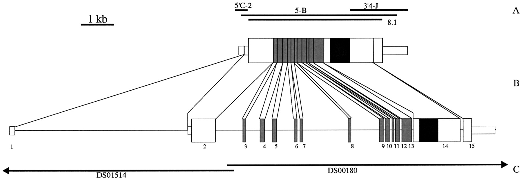
Figure 1.
Schematic representation of the DLGR-2 cDNA and genomic clones and the organization of the DLGR-2 gene. (A) Positions of the PCR clones. (B) Schematic drawing of the composite cDNA (top) and the organization of the receptor gene (bottom). The exons are given as bars and numbered 1–15. We named the introns after the preceeding exons(e.g., intron 1 follows exon 1). The narrow and broad bars represent noncoding and coding regions, respectively. The DNA region coding for the transmembrane domain is black and that coding for the Leu-rich repeats are gray. (C) The positions of the genomic P1 clones, DS00180 and DS01514, from the Berkeley DrosophilaGenome Project.


