
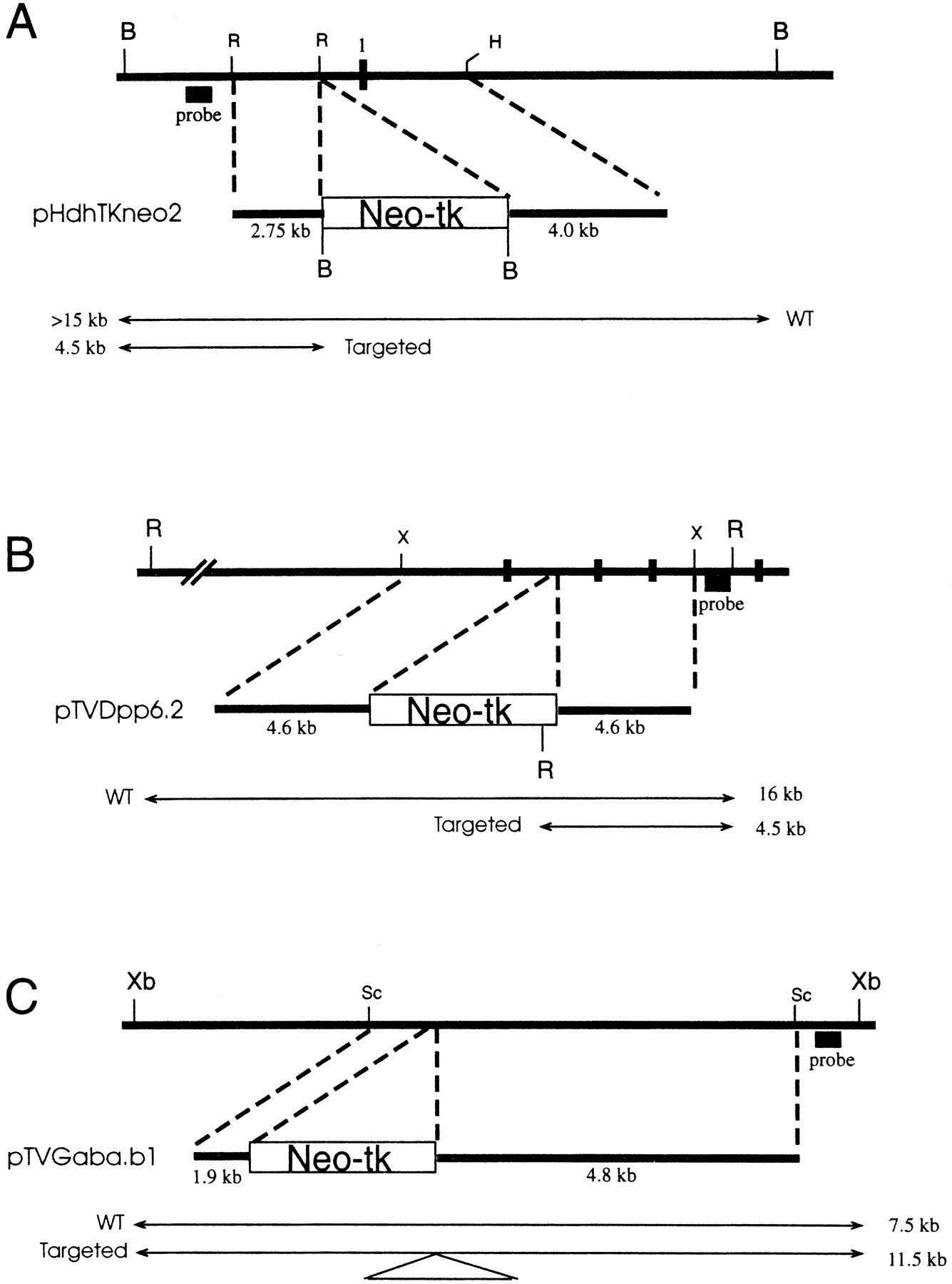
Targeting vectors. (A) Hdh targeting vector. Transcriptional orientation of the gene is to the right. Note position of exon 1 (1). (B) Dpp6 targeting vector. (C) Gabrb1 targeting vector. In each panel, the genomic locus is shown at the top as a horizontal thick line. Positions of exons are indicated by vertical broken bars. Probes used to detect targeting events are indicated, and the sizes of endogenous (WT) and targeted restriction fragments are shown next to arrows spanning the endpoints. The names of targeting vectors are shown, with the location of the HSV-tk (tk)/neomycin resistance (neo) cassette indicated in the large rectangular boxes. (B) BamHI; (R ) EcoRI; (H) HindIII; (X or Xb) XbaI; (Sc) SacI.


