
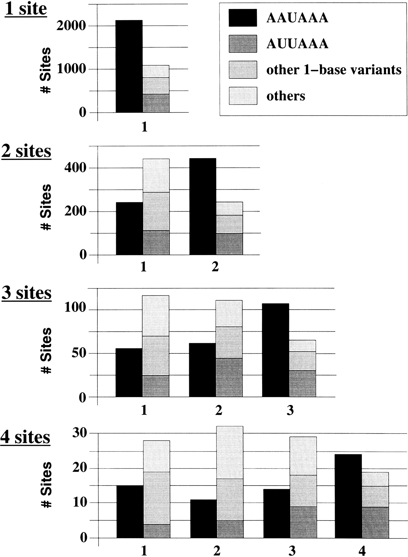
Figure 2.
Distribution of polyadenylation signal types at each site on the UTR. From top to bottom: mRNAs with a single poly(A) site identified, mRNAs with two poly(A) sites identified, mRNAs with three poly(A) sites identified, and mRNAs with four poly(A) sites identified. Poly(A) sites are numbered from 5′ to 3′.


