
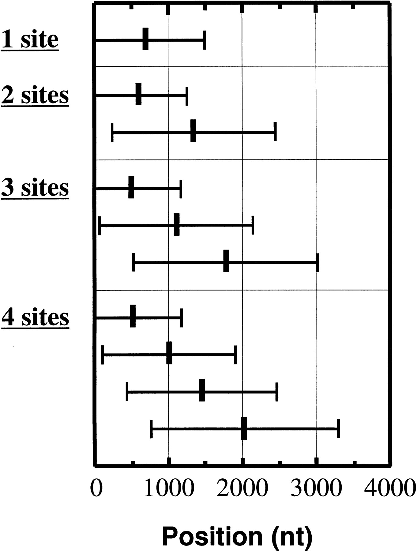
Figure 1.
Average position of observed polyadenylation sites on 3′ UTRs in function of the number of observed alternate sites. From top to bottom: mRNAs with a single poly(A) site identified (3377 RNAs), mRNAs with two poly(A) sites identified (724 RNAs), mRNAs with three poly(A) sites identified (182 RNAs), and mRNAs with four poly(A) sites identified (43 RNAs). Position 1 on the X-axis is the first base following the Stop codon. Error bars indicate standard deviations.


