
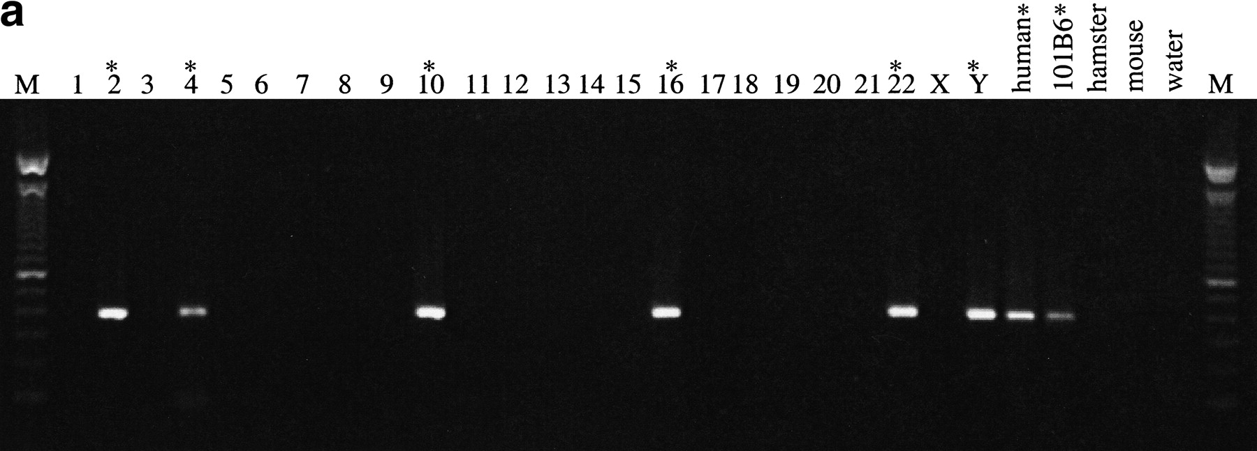
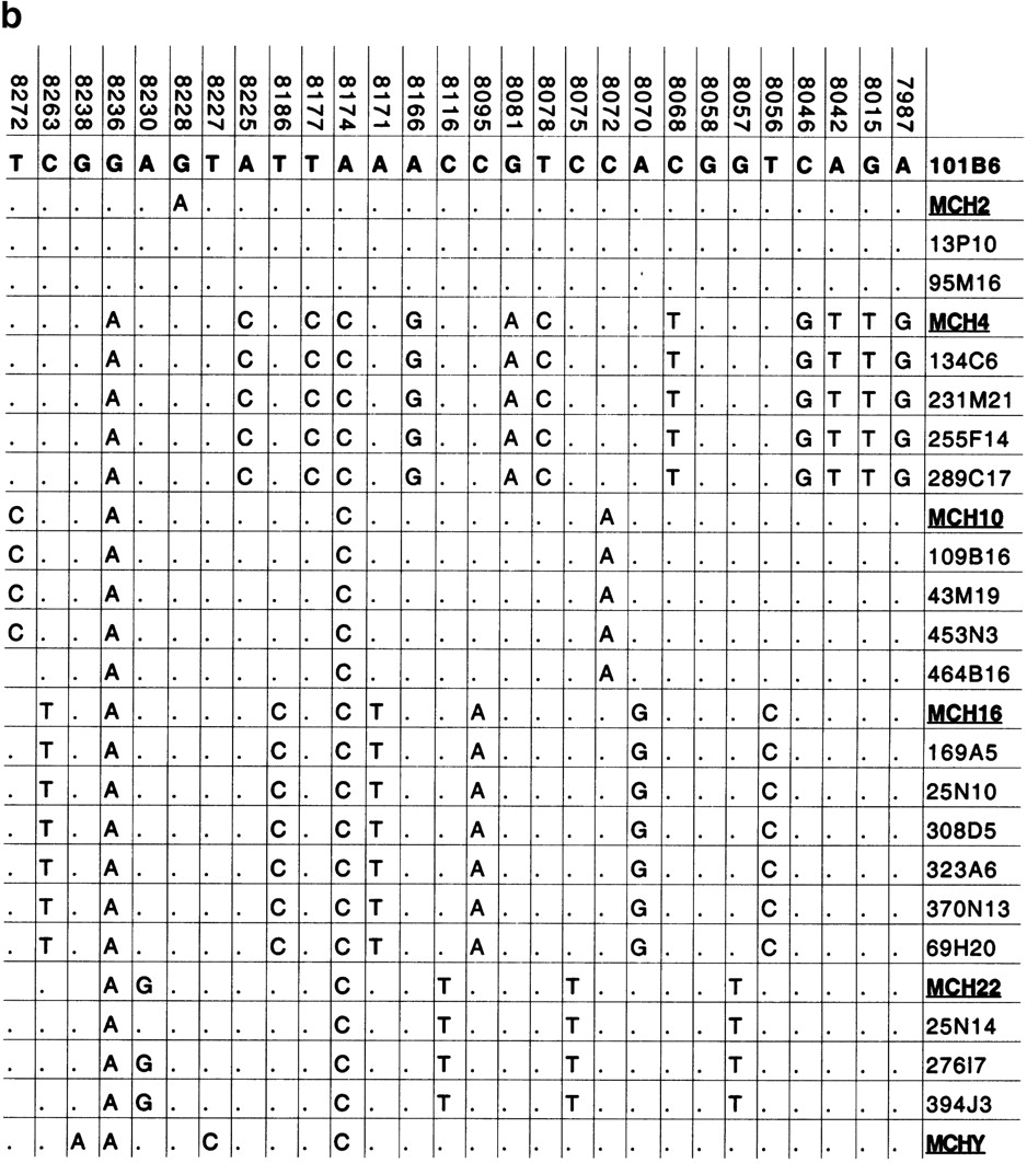
Paralogous STS and sequence variants. (a) A typical PCR amplification of a paralogous STS against a panel of monochromosomal somatic cell hybrid DNAs. pSTS1 was designed to 101B6 (chromosome 2) sequence (see Methods) yet amplified a ∼383 bp product from chromosomes 2, 4, 10, 16, 22, and Y (marked with asterisks). (b) The PCR products from pSTS 1 were bidirectionally sequenced and aligned (Consed). Basepairs in boldrepresent 101B6 basepairs, whereas the numbers above each bp represent its location in 101B6. Only the paralogous sequence variants (PSVs) that distinguish each chromosome are shown; a periodrepresents the same bp as 101B6. Along the right are the sequences of the monochromosomal hybrid sequence (MCH). Below each chromosomal sequence signature, a subset of RPCI-11 BAC clones corresponding to each PSV is indicated. The numbers correspond to pSTSs developed to the 101B6 reference sequence. Similar analyses were performed for 16 other pSTS.


