
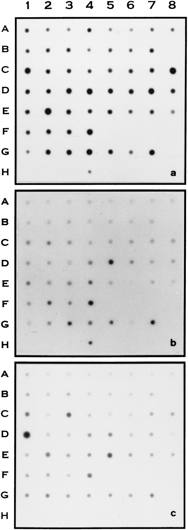
RNA master blot analysis of CGI genes in human tissues. Master tissue blots were hybridized with cloned RT-PCR-amplified fragments of human CGI genes as indicated. (a) CGI-7. (b) CGI-17. (c) CGI-27. The exposure time for each blot was 3 days for A, 7 days for B and 20 hours for C. The tissue distribution on the blot from left to right in order (1–8) was: A, whole brain; amygdala; caudate nucleus; cerebellum; cerebral cortex; frontal lobe; hippocampus; medulla oblongata. B, occipital pole; putamen; substantia nigra; temporal lobe; thalamus; subthalamic; nucleus; spinal cord. C, heart; aorta; skeletal muscle; colon; bladder; uterus; prostate; stomach. D, testis; ovary; pancreas; pituitary gland; adrenal gland; thyroid gland; salivary gland; mammary gland. E, kidney; liver; small intestine; spleen; thymus; peripheral leukocyte; lymph node; bone marrow. F, appendix, lung, trachea, placenta. G, fetal brain; fetal heart; fetal kidney; fetal liver; fetal spleen; fetal thymus; fetal lung. H, yeast total RNA; yeast tRNA; E. coli rRNA; E. coli DNA; Poly r(A); human C0t DNA; human DNA; human DNA.


