
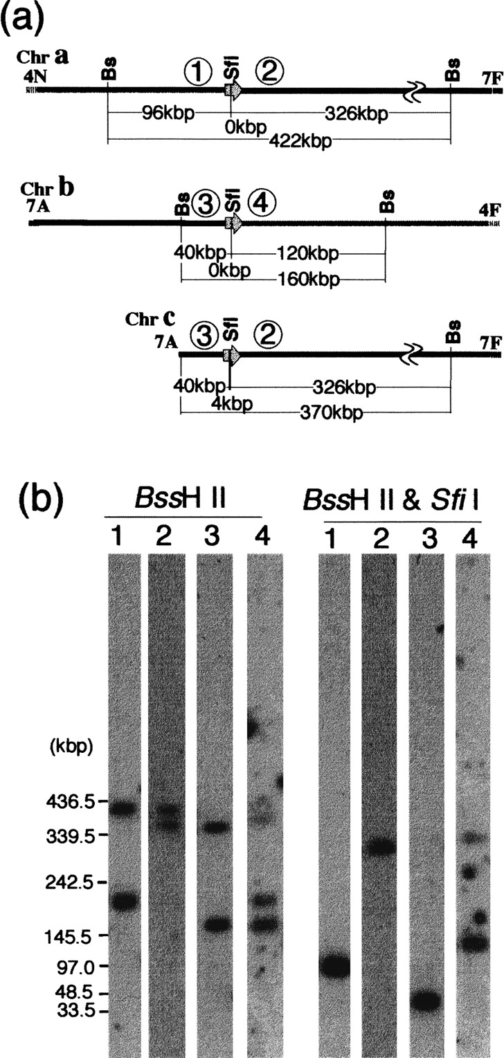
Junction of the translocation between chromosomes 4 and 7 in NUM1000.BssHII and SfiI restriction maps of the junction (a). The locations of the MRS flanking markers are indicated by numbers in circles: (1) OGR4; (2) DBP7; (3) YPL12; and (4) OGH12. The locations of YPL12 and DPB7 in chromosome 7 are shown in Figure 1. OGR4 and OGH12 were assigned to SfiI fragments 4N and 4F of chromosome 4, respectively (not shown). Southern hybridization with MRS flanking markers of chromosomes 4 and 7 (b). Genomic DNA of WO-1 was digested with BssHII and withBssHII and SfiI. The restriction fragments were separated by contour-clamped homogeneous electric field. The fragments were then transferred onto membranes and hybridized with OGR4, lane1; DBP7, lane 2; YPL12, lane3; and OGH12, lane 4. The DNA size was determined by Lambda ladder DNA markers.


