
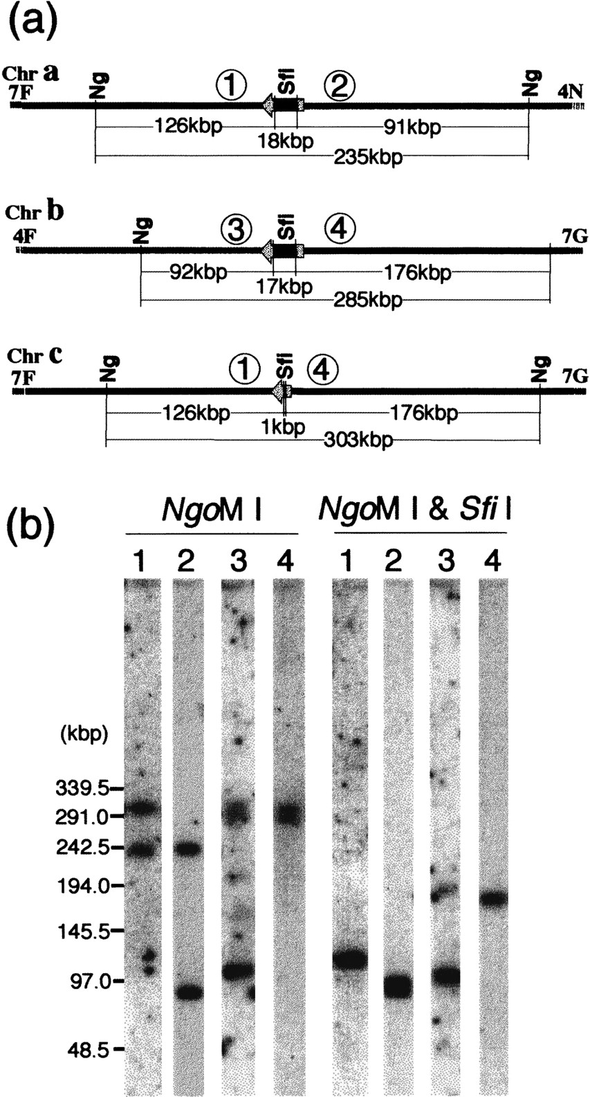
Junction of the translocation between chromosomes 4 and 7 in WO-1.NgoMI and SfiI restriction map of the junction (a). The locations of the MRS flanking markers are indicated by numbers in circles: (1) RBP1; (2) OGR4; (3) OGH12; and (4) ARS3. The locations of RBP1 and ARS3 in chromosome 7 are shown in Figure 1. OGR4 and OGH12 were assigned to SfiI fragments 4N and 4F of chromosome 4, respectively (not shown). Southern hybridization with markers flanking the MRSs of chromosomes 4 and 7 (b). Genomic DNA of WO-1 was digested with NgoMI and withNgoMI and SfiI and resolved as described in Methods. The fragments were then transferred onto membranes and hybridized withRBP1, lane 1; OGR4, lane 2; OGH12, lane3; and ARS3, lane 4. OGH12 shows weak cross-hybridizaton to 7F. The DNA size was determined by Lambda ladder DNA markers.


