
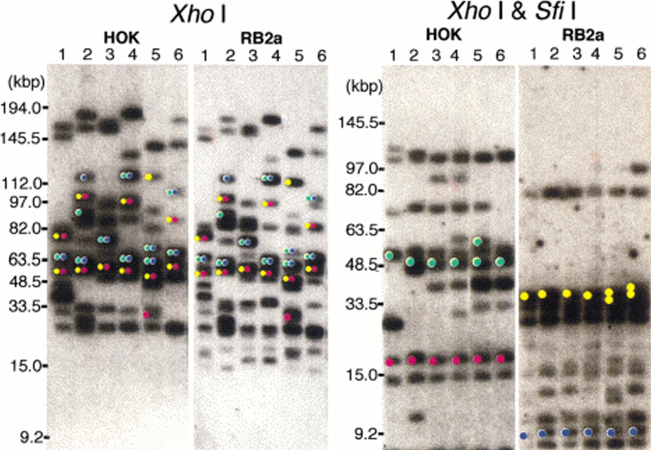
Figure 4.
Orientation of the MRS with respect to the flanking markers. The membranes shown in Figure 3 were stripped and rehybridized with RB2a (a part of RB2) and HOK. The bands shown to hybridize to the flanking markers are indicated by colored circles. The bands hybridized to the MRS flanking markers YPL12 (yellow), DBP7 (red),RBP1 (green), and ARS3 (blue) in Figure 3 indicated by dots on the hybridization profiles.


