
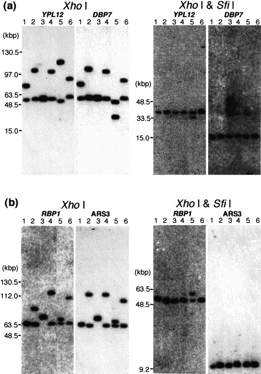
Southern hybridization with probes flanking the MRS to restriction fragments of XhoI and XhoI–SfiI digests. Because the MRSs in chromosome 7 do not include XhoI sites, aXhoI digest leaves fragments including the entire MRS sequences and the adjacent DNA. Four MRS flanking markers,YPL12, DBP7, RBP1, and ARS3 were included in the XhoI fragments (Fig. 1). The genomic DNA of each strain was digested with XhoI and with XhoI andSfiI. The DNA was separated by countour-clamped homogeneous electric field and blotted onto a membrane. Lane 1, 1006; lane2, WO-1; lane 3, NUM55; lane 4, NUM114; lane5, NUM1000; lane 6, 1719. The DNA size was determined with the MidRange 1 PFG marker (New England Biolabs) and 1 kb ladder (Bethesda Research Laboratories). The MRS between 7A and 7F: probesYPL12 and DBP7 (a). The MRS between 7F and 7G: probesRBP1 and ARS3 (b).


