
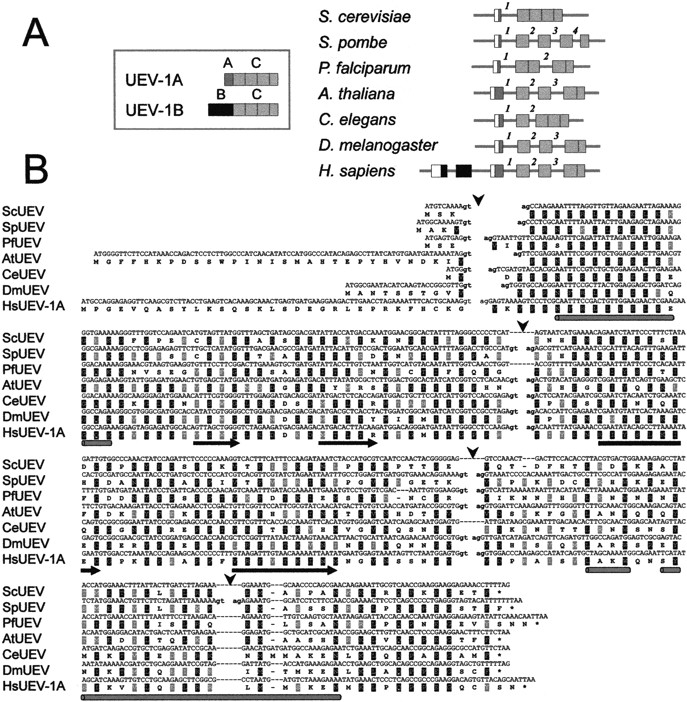
Conservation of introns in UEV genes from distant organisms. (A) Diagrammatic representation (not to scale) of the relative exon–intron arrangement of the UEV gene in Saccharomyces cerevisiae, Schizosaccharomyces pombe, P. falciparum, Arabidopsis thaliana, Caenorhabditis elegans, Drosophila melanogaster, and Homo sapiens. Inset diagram for the two major isoforms described for human UEV1. Putative 5′ untranslated (UTR) segments are represented as open boxes. Introns are designated by numbers that are specific for each UEV gene. Vertical dotted lines within exons define segments corresponding to the exons in S. pombe UEV. (B) Alignment of UEV genes and proteins showing precise conservation of the position of the second and third introns interrupting the C domain of UEV proteins. Splice donor and acceptor sequences in intron boundaries are in lower case. The positions of introns are marked by arrowheads. Predictions of secondary structure are shown below the sequences as rods (helices) or arrows (strands).


