
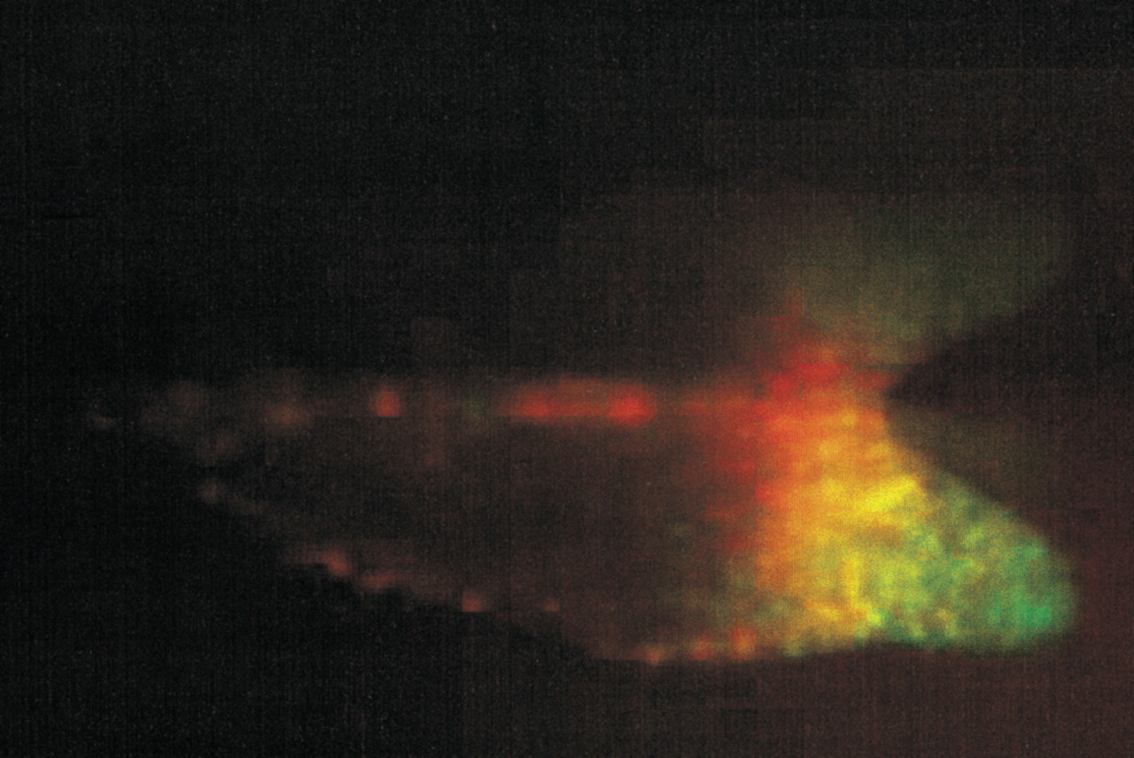
This is a living Dictyostelium slug containing ∼20,000 prestalk cells in the front one-fifth and 80,000 prespore cells in the rear four-fifths. There are also scattered prestalk-like cells in the prespore region. There are two kinds of prestalk cells in the prestalk region that are defined by their use of different promoter elements from the ecmA gene. In the slug shown in this image (D. Dorman, T. Abe, J. Williams, and C. Weijer, unpubl.) the two promoter regions were coupled to different GFPs and the fluorescence is from pstA cells (red) in the tip, pstO cells (green) behind them, and a region where the two cell populations overlap (yellow).


