
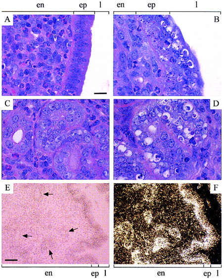
Histology (A–D) and in situ hybridization (E,F) of uterine sections. (A,C) Ten-week-old, hematoxylin-and eosin-stained C57+/+ uterus (day 0.5 VP), showing normal morphology of the endometrial epithelium lining the uterine lumen (A) and endometrial glands (C). (B,D) ten-week-old, hematoxylin- and eosin-stained C57−/− uterus (day 0.5 VP), showing highly disorganized and apoptotic (clear) epithelial cells lining the uterine lumen (B) and endometrial glands (D). Apoptosis of the clear cells (containing condensing chromatin) was confirmed by TUNEL assay (Gavrieli et al. 1992) (data not shown). Scale bar for A–D, 20 μm. (E,F) Bright-field (hematoxylin-stained) and dark-field views, respectively, of 10-week-old R1+/+ uterus hybridized with a35S-labeled mouse Cenpb antisense riboprobe, showing mRNA signals (dark brown grains in E and white grains in F) throughout the endometrium (and the myometrium; not included in picture) with maximal mRNA expression in the epithelial lining of the uterine lumen and endometrial glands (selected examples of which are indicated by arrows). Scale bar for E and F, 50 μm. (en) Endometrium; (ep) epithelium, (l) uterine lumen.


