
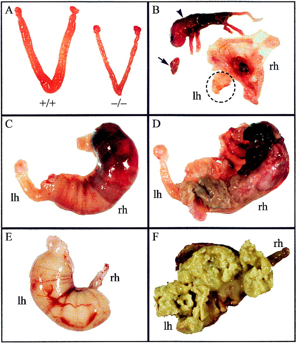
Uterine and pregnancy problems in Cenpb null mice. (A) Size comparison of uteri from 10-week-old R1+/+and R1−/− mice, showing the left and right uterine horns. The sizes of the ovaries and oviducts attached to these horns were normal in both animals. (B) Nine-month-old, 1-week overdue W9.5−/− pregnant female (first pregnancy), showing a full-term dead fetus (arrowhead) attributable to placental necrosis in the right horn (rh) and fetal growth arrest/resorption (arrow) in the left horn (lh; shown in circle). (C,D) Twelve-month-old, 10-day-overdue R1−/− pregnant female (fourth pregnancy), showing external and internal views of a necrotic fetus in the right horn, and absence of fetuses in the left horn. (E,F) Nine-month-old, 4-day-overdue R1−/− pregnant female (first pregnancy), showing external and internal views of decomposed fetal content in the left horn, and absence of fetuses in the right horn.


