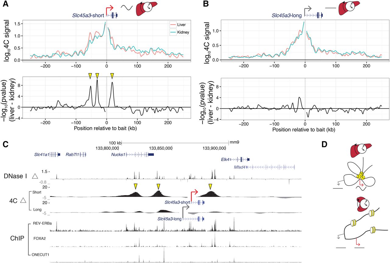
Precise promoter-enhancer contacts underlie liver-specific mRNA rhythms. (A,B) 4C-seq profiles for the (A) Slc45a3-short and (B) Slc45a3-long isoforms within ±250 kb around baits targeting the two TSSs (top). Signed log P-values for differential contacts between liver and kidney (bottom) as in Figure 5B. TSSs for Slc45a3-short and Slc45a3-long are 8 kb apart. (C) Differential 4C contacts (signed log P-values), log2 fold change of DNase I hypersensitivity between liver and kidney, and ChIP-exo signal of REV-ERBa, FOXA2, and ONECUT1. Regions of significant differential contacts in Slc45a3-short correspond to liver-specific DNase I hypersensitive regions. Yellow arrowheads in A and C show liver-specific distal contacts recruited to the Slc45a3-short TSS. These contacts are absent for Slc45a3-long TSS (B). (D) Schematic model illustrating enhancer-promoter interactions in liver and kidney that may generate liver-specific rhythms. Yellow circles illustrate liver-active enhancers contacting the rhythmic promoter (red arrow) but not the alternative nonrhythmic promoter (gray arrow). In kidney, the enhancer is not accessible, and both promoters are nonrhythmic.