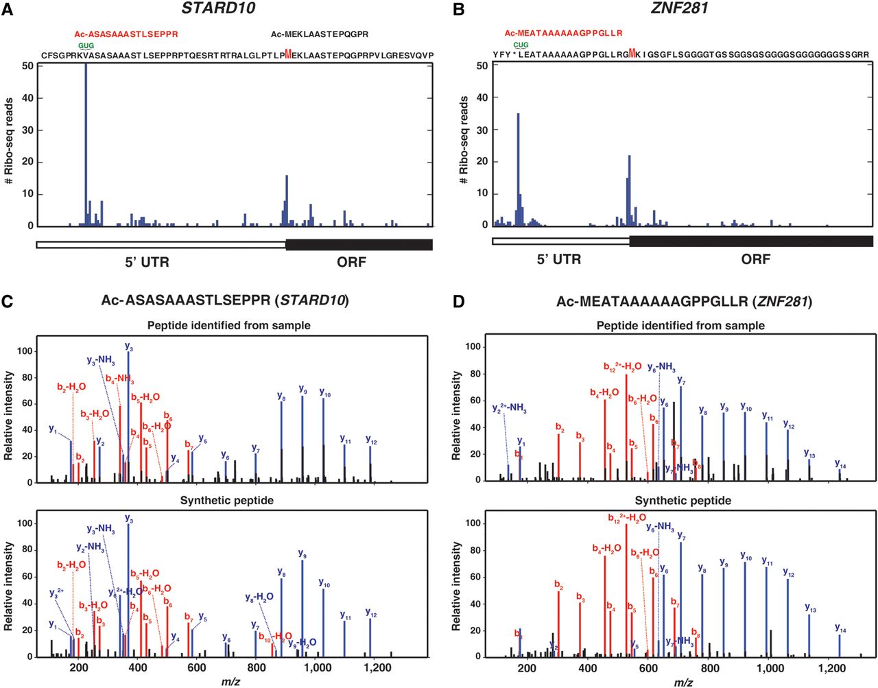
Acetylated peptides identified in 5′ UTRs map to TISs inferred using ribosome profiling data and were validated by synthetic peptides. (A) The acetylated peptide identified from HEK293T cells that was derived from the 5′ UTR of STARD10 along with the annotated TIS (the methionine “M” is marked in red) were aligned with ribosome profiling data from HEK293 cells. (B) The acetylated peptide identified from HEK293T cells that was derived from the 5′ UTR of ZNF281 was aligned with the ribosome profiling data from HEK293 cells. The methionine of the annotated TIS is marked as “M” in red. (C) The acetylated peptide positioned in the 5′ UTR of STARD10 was validated with synthetic peptides. The annotated mass spectrum derived from the sample (top) is aligned with the mass spectrum derived from a synthetic peptide (bottom). (D) The acetylated peptide positioned in the 5′ UTR of ZNF281 was validated with synthetic peptides. The annotated mass spectrum derived from the sample (top) is aligned with the mass spectrum derived from a synthetic peptide (bottom).