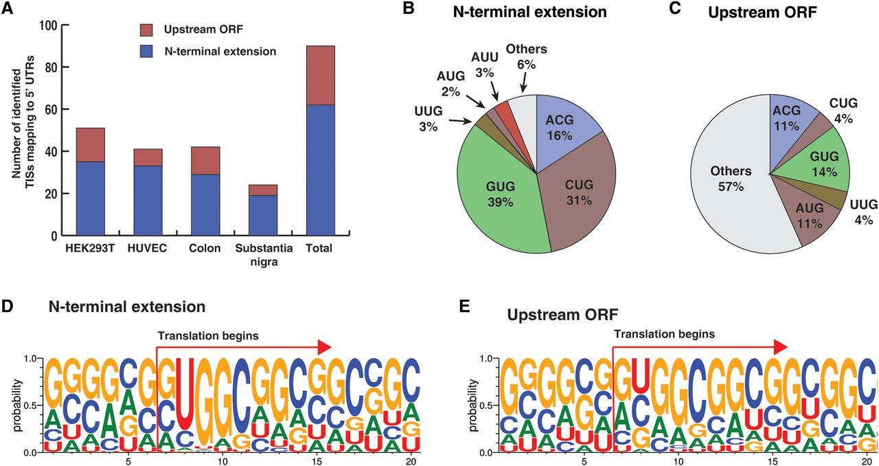
Translation initiation sites mapping to 5′ UTRs. (A) Peptides acetylated at their N termini mapping to 5′ UTRs that were identified from HEK293T cells, HUVEC, human colon, and human substantia nigra as shown. The colors indicate whether the peptides correspond to uORFs or were N-terminal extensions of annotated proteins. (B) Codons for TISs corresponding to acetylated peptides mapping to 5′ UTRs which led to N-terminal extension of annotated proteins. (C) Codons for TISs corresponding to acetylated peptides mapping to 5′ UTRs which encode an uORF. (D) Sequence logo of nucleotides surrounding the TIS in cases where the TISs lead to N-terminal extension of annotated proteins. (E) Sequence logo of nucleotides surrounding the TIS in cases where the TISs are located in an uORF.