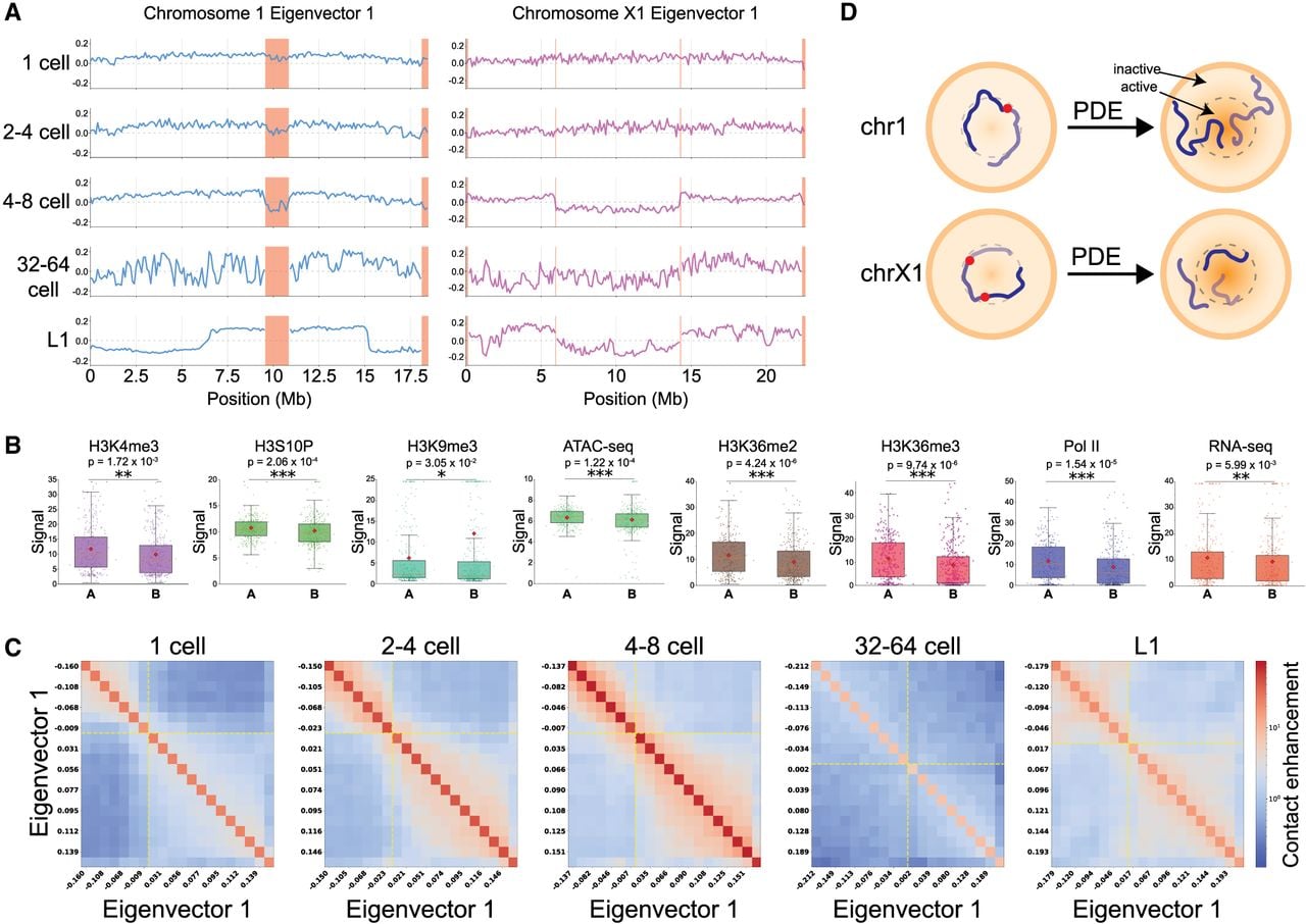
PDE splits chromosomes and causes 3D genome changes. (A) Compartmentalization changes during Ascaris embryogenesis. Hi-C compartments were determined by using the eigenvector 1 values through the developmental stage. Eliminated regions are highlighted in light red (for compartments for all chromosomes, see Supplemental Fig. S7). For Chr 1, weak compartmentalization between its two future somatic derivatives is shown before PDE. Following PDE, the 3′ end of Chr 1a and the 5′ end of Chr 1b occupy the same active compartment, and the 5′ end of Chr 1a and the 3′ end of Chr 1b remain in the inactive compartment (see also D). For Chr X1, after PDE split it into three somatic chromosomes, the two terminal fragments occupy the same compartment, and the central fragment is segregated away (see also D). (B) Multiomic analysis confirms the defined compartments. Active chromatin marks are enriched in A compartments, and a heterochromatin mark is enriched in B compartments. Each point represents a 100 kb genomic region. A two-sided Mann–Whitney U-test was performed to calculate the statistical significance for each comparison: (***) P < 0.001, (**) P < 0.01, and (*) P < 0.05. (C) Dynamics of compartment interactions through development. A/B-compartment interactions are shown using saddle plots. Genomic bins are sorted by PC1 value and binned into 20 quantiles (both axes). Contact enrichment within each bin pair is displayed (red = high; blue = low). Dashed yellow lines mark the A/B boundary (PC1 = 0). (D) Model of chromosomal rearrangements following PDE. The germline chromosomes are not strongly compartmentalized in the early embryos. After PDE, the resulting somatic chromosomes reorganize their compartments. The two chromosomes were modeled based on data from A.