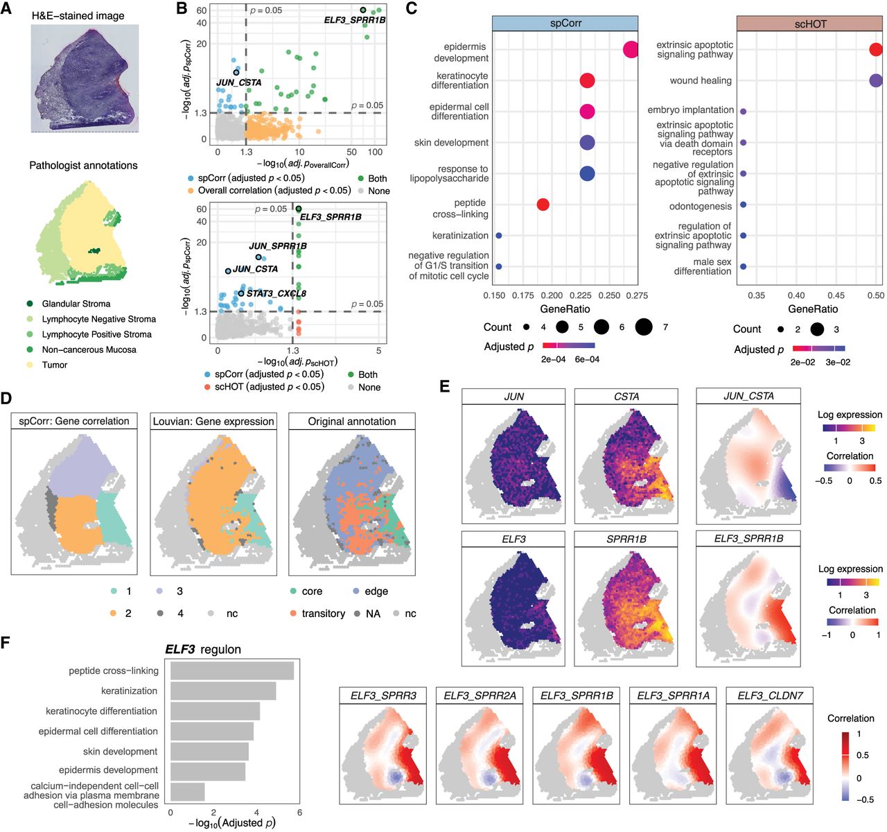
spCorr elucidates intratumoral regulatory heterogeneity. (A) H&E-stained image and pathologist-defined spatial annotations for sample 2 of the HPV-negative OSCC data set. The tumor region is distinguished from the surrounding stromal and mucosal tissue. (B) Scatter plots comparing adjusted p-values from spCorr with those from non-spatial overall correlation (top) and scHOT (bottom) across all candidate TF–target gene pairs. Colors indicate whether a gene pair is identified as significant (BH-adjusted p < 0.05) by spCorr only, by the alternative method only, by both methods, or by neither. (C) GO enrichment analysis of genes identified by two methods. spCorr-identified genes are enriched for epidermis- and keratinocyte-related processes consistent with OSCC biology, whereas genes identified by scHOT show broader and less specific enrichment patterns. (D) Left: Spatial clustering based on spCorr-inferred local correlation estimates reveals tumor subdomains that aligned with original annotations (right) while offering smoother and more clearly defined boundaries. Middle: Louvain clustering based on expression of the same TF-target gene pairs fails to produce comparably distinct boundaries. (E) Spot-level correlation estimates for two representative TF-target pairs. Top: the JUN and CSTA pair exhibits a negative correlation in the tumor core and a positive correlation in the leading edge and transitory region, suggesting localized inflammatory activity in tumor peripheral areas. Bottom: the ELF3 and SPRR1B pair displays a strong positive correlation in the tumor core, consistent with ELF3’s known role in epithelial differentiation. (F) Functional analysis of the ELF3 regulon. Left: GO enrichment analysis of ELF3 regulon reveals biological processes including keratinization and epidermal development. Right: local correlation estimates for ELF3 and its targets are aligned with the boundary of the tumor core, indicating their domain-specific transcriptional activities.