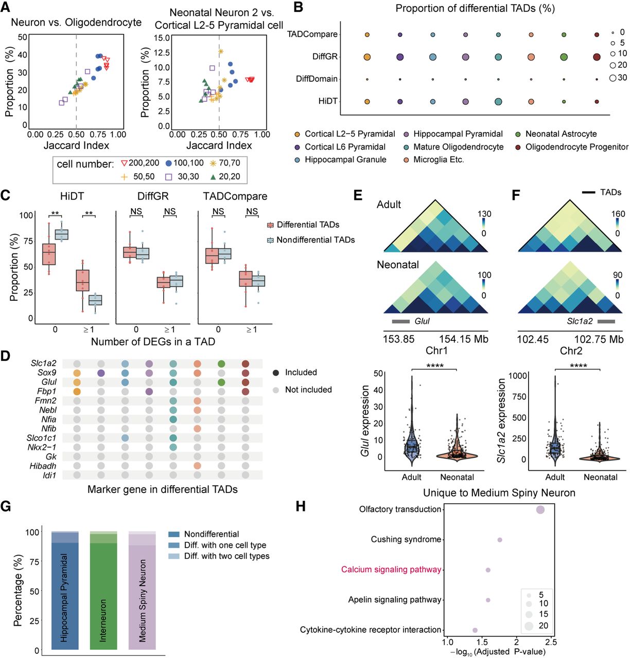
Identification of differential TADs across cell subtypes from mouse brain single-cell Hi-C data. (A) Proportions and Jaccard indices of differential TADs identified from increasing numbers of sampled cells for neurons versus oligodendrocytes and neonatal neuron 2 versus cortical L2–5 pyramidal cells. Five replicates are shown for each cell number, and the dashed line marks a Jaccard index of 0.5. (B) Proportions of differential TADs identified across brain cell types. (C) Proportions of differential and nondifferential TADs containing no DEGs or at least one DEG. Statistical significance was assessed using a Wilcoxon signed-rank test: (**) P < 0.01. (D) Astrocyte marker genes that are located within differential TADs. (E,F) Differential TADs containing Glul and Slc1a2 between adult and neonatal astrocytes, with corresponding single-cell expression levels. Statistical significance was evaluated using a two-sided paired t-test: (****) P < 0.0001. (G) Proportion of differential TADs identified among hippocampal pyramidal, interneurons, and medium spiny neurons. Differential TADs are categorized as being specific to one subtype or shared across two subtypes. (H) GO enrichment of genes within differential TADs shared between the medium spiny neuron comparisons.