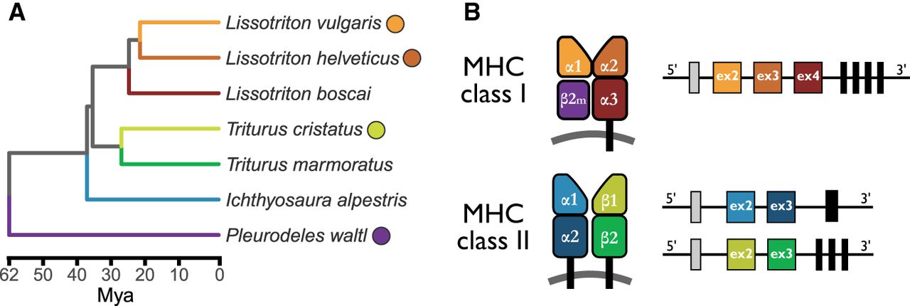
Newt phylogeny and a schematic representation of MHC genes and proteins. (A) Time-calibrated phylogeny of the studied species (Stewart and Wiens 2025); colored dots next to the species name indicate the availability of a reference genome. (B) Schematic representation of the structure of MHC molecules and their genes. MHC-I molecules are composed of a heavy chain with three extracellular domains (α1, α2, and α3), paired with a nonvariable β2-microglobulin (β2m) molecule, which is encoded by a gene outside the MHC region. MHC-II molecules are heterodimers, each consisting of an α and a β chain. Each chain contributes two extracellular domains: α1 and α2 for the α chain, and β1 and β2 for the β chain. The peptide-binding groove in MHC-I is formed by the α1 and α2 domains, whereas in MHC-II it is formed by the α1 and β1 domains. All MHC molecules also include a transmembrane domain and a cytosolic tail. There is a direct correspondence between structural domains and exons, as illustrated on the right-hand side. The first exon (gray box) encodes a leader peptide; each extracellular domain is encoded by a separate exon (indicated as “ex” with the exon number inside the box). The remaining regions—including the transmembrane domain and cytosolic tail—are encoded by a variable number of exons (black boxes), typically ranging from one to four, depending on the molecule type.