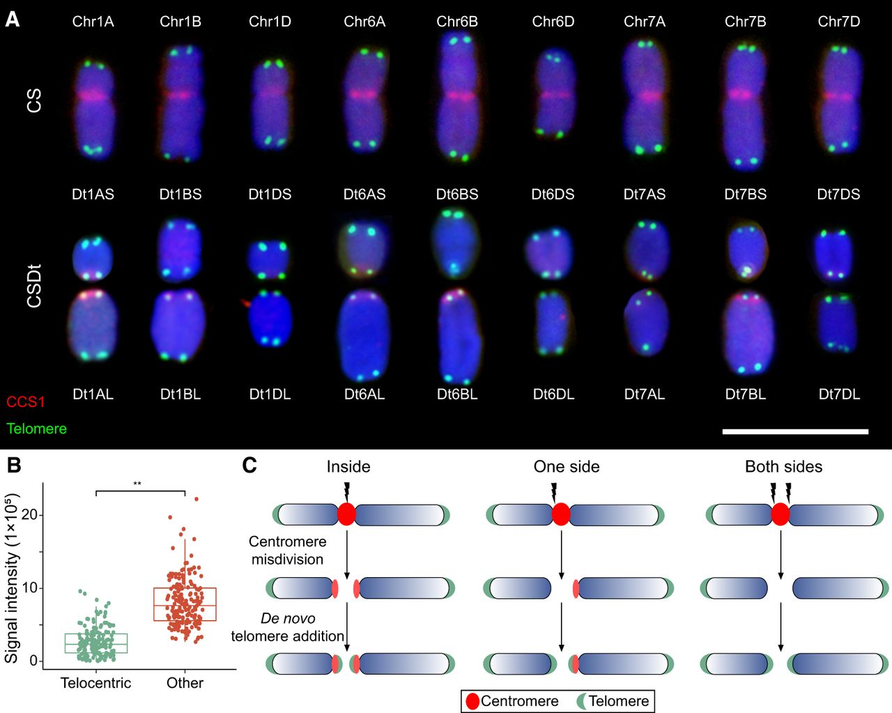
Distribution of centromere and telomere signals on ditelosomic and corresponding chromosomes in Chinese Spring (CS) wheat. (A) Fluorescence in situ hybridization (FISH) analysis illustrates centromere and telomere distribution on ditelosomic chromosomes derived from homoeologous groups 1, 6, and 7, as well as on their corresponding full-length chromosomes in Chinese Spring wheat. Chromosomal DNA is counterstained with 4′,6-diamidino-2-phenylindole (DAPI, blue). Centromeres are labeled in red using the centromere-specific probe CCS1, and telomeres are labeled in green using a telomere-specific probe. Each panel displays the short arm (S) and long arm (L) telosomes from A, B, and D genomes, along with the full chromosomes (Chr 1A–Chr 1D, Chr 6A–Chr 7D), clearly showing variations in centromere and telomere positioning and signaling. Scale bar is 10 µm. (B) Fluorescence intensity of CRW fluorescence in long-arm ditelosomic lines. Centromeric signals from two telocentric chromosomes were compared with those from other intact chromosomes for each line. The y-axis indicates relative CRW fluorescence intensity, with error bars representing standard deviation (s.d.). (**) Significant differences at P < 0.01 (Student's t-test). (C) A schematic model illustrating centromere misdivision-induced chromosome breakage in wheat. Three scenarios of centromere breakage are depicted: breakage within centromere (internal), on one side, or on both sides of the centromere. Chromosome healing is hypothesized to occur through the de novo addition of telomeric repeats at the breakpoint ends. Chromosome arms are shown in blue, centromeres in red, and telomeres in green.