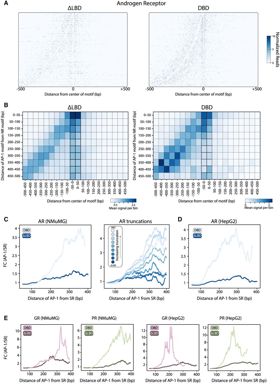
Binding of the SRDBD mutants is stronger at the proximal AP-1 motif than at the SR motif itself. (A) Visualizing the signal-footprints of adjacent motifs. Genomic regions from the peaks containing both SR (canonical) and AP-1 motifs (noncanonical) were aligned to the SR motif center and sorted by intermotif distance, with AP-1 positioned upstream for ARΔLBD (left) and ARDBD (right). The heatmap shows MNase-cleaved fragment ends, revealing protection patterns at both motif sites. (B–E) Proximity to AP-1 motif increases binding of ARDBD to its canonical motif. (B) The bindings of ARΔLBD (left) and ARDBD (right) binned by distance from SR center (x-axis, 50 bp) and AP1-to-SR spacing (y-axis, 50 bp). The color scale shows mean normalized reads per bin. (C, left) The distance-dependent effect is summarized, showing the fold-change of the signal at the AP-1 versus SR motifs as a function of the distances between motifs. The same analysis is also shown for the set of AR truncations (C, right) and for two mutants of the AR in the HepG2 cell line (D). (E) The distance-dependent effect for the GR and PR mutants in NMuMG and HepG2 cell lines.